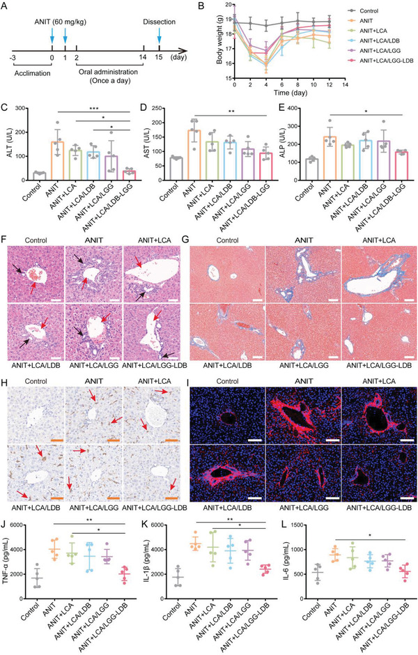
Figure 2.

Amelioration of ANIT‐induced acute liver bile duct injury in mouse model. A) The therapeutic process of ANIT‐induced mice model of cholestasis and acute liver bile duct injury. B) The changes of mice body weight during treatment. Liver function‐related enzymes concentration of C) ALT, D) AST, and E) ALP. F) H&E staining of liver tissues in different treatment groups (Red arrows: interlobular vein (IV); Black arrows: interlobular bile duct (IB); Scale bar: 50 µm). G) Hepatic Masson staining in different treatment groups (Scale bar: 100 µm). H) Hepatic immunohistochemical staining of F4/80 marker‐positive macrophages (red arrows) in different treatment groups (Scale bar: 50 µm). I) Hepatic immunofluorescence staining of collagen‐I in different groups (Scale bar: 100 µm). The hepatic inflammatory factors of J) TNF‐α, K) IL‐1β, L) IL‐6. The data were presented as the mean ± s.d., n = 5. The statistical significance was calculated via one‐way ANOVA with Tukey's multiple comparisons test. *P < 0.05, **P < 0.01, ***P < 0.001.
