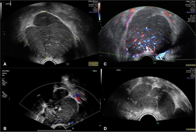
Figure 1.
(A) 2D grey scale ultrasound image of the solid lesion of the right adnexa. (B) 2D colour ultrasound image of the solid lesion of the right adnexa with acoustic shadows. (C) 2D colour doppler ultrasound image of the solid lesion of the right adnexa with moderate blood flow (colour score 3). (D) Ultrasound image of the solid lesion attached to the left ovary with the same features.

