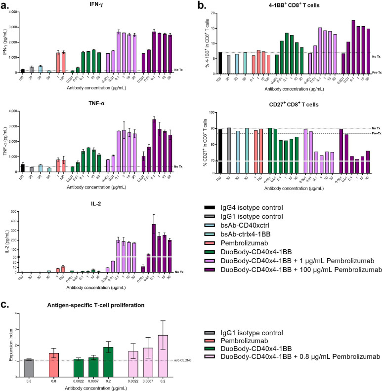
Figure 6.
Combination of DuoBody-CD40×4-1BB with pembrolizumab amplifies the magnitude of the immune response. (A, B) Purified CD8+ T cells from healthy donors were cocultured with LPS-matured allogeneic DCs in the presence of DuoBody-CD40×4-1BB, research-grade pembrolizumab (either alone or in combination (concurrent treatment)) or control antibodies for 5 days. (A) Cytokine concentrations in supernatant taken after 5 days of culture in the presence of indicated antibodies. Data shown are mean concentration±SD of duplicate wells from one representative donor pair (donor pair two is shown in online supplemental figure 5). Dashed line indicates cocultures that were not treated with antibody (No Tx). (B) The percentage of 4-1BB+ CD8+ T cells and CD27+ CD8+ T cells was measured by flow cytometry. Data shown are the percentage of positive cells within the total CD8+ T-cell population from one representative donor pair (donor pair two is shown in online supplemental figure 5). Dashed line indicates cocultures on Day 0 (before antibodies were added to the treated cocultures) and cocultures that were not treated with antibody at Day 5 of the MLR assay (No Tx). (C) CD8+ T cells were electroporated with RNA encoding an HLA-A2/CLDN6-specific TCR and PD-1, labeled with CFSE and cocultured with autologous DCs electroporated with CLDN6-encoding RNA in presence of the DuoBody-CD40×4-1BB (0.0022, 0.0067 or 0.2 µg/mL), IgG1 isotype control (0.8 µg/mL), clinical-grade pembrolizumab (0.8 µg/mL) or combinations thereof for 5 days. CFSE dilution in CD8+ T cells was analyzed by flow cytometry. Mean expansion index±SD (n=4) is shown. Dashed line indicates CD8+ T cells that were cocultured with DCs that were not electroporated with CLDN6 in the presence of the IgG1 isotype control antibody as negative control. CFSE, carboxyfluorescein succinimidyl ester; DC, dendritic cell; LPS, lipopolysaccharide; TCR, T-cell receptor.

