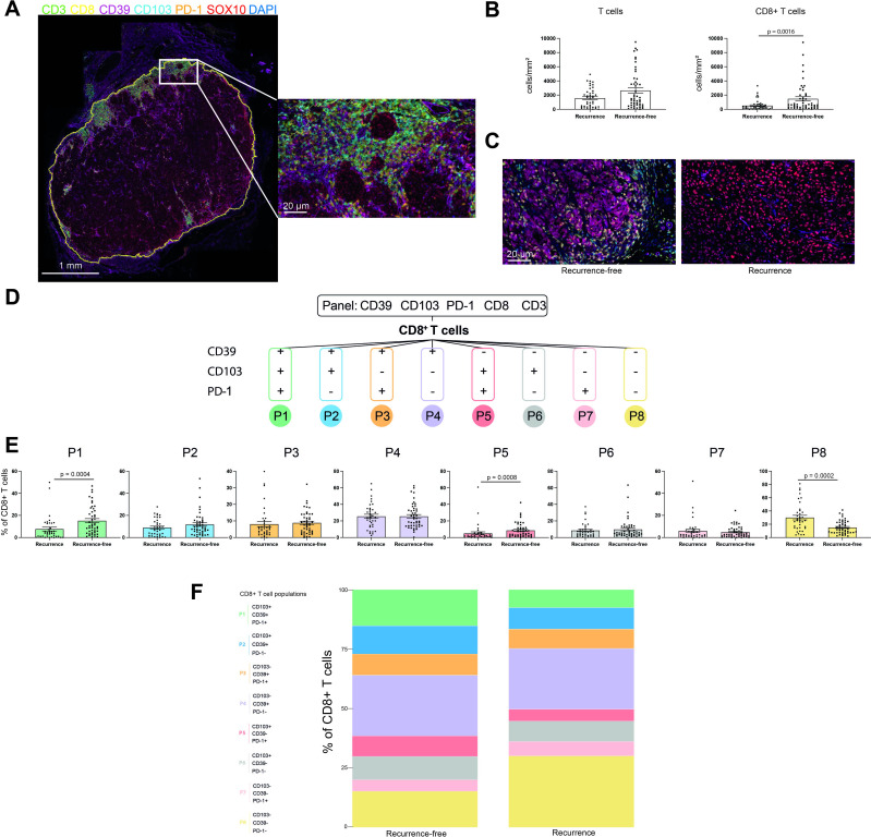
Figure 1.
mIHC identifies significant CD8+ T-cell populations in adjuvant PD-1-treated patients with stage III melanoma. (A) mIHC was performed on pre-treatment stage III melanoma FFPE tissue from patients receiving adjuvant anti-PD-1 immunotherapy. Tumors were stained for CD3 (green), CD8 (yellow), CD39 (magenta), CD103 (cyan), PD-1 (orange), SOX10 (red) and DAPI (blue). Analysis was limited to intratumoral regions of tissue (highlighted in yellow). (B) Intratumoral T cells and CD8+ T cells CD8+ T cells were quantified per square millimetre of tumor and compared between recurrence patients and recurrence-free patients. Statistical differences were calculated using a non-parametric Mann-Whitney test (n=91). (C) Representative mIHC-stained FFPE sections from an RF patient and an R patient. (D) CD8+ T cells were divided into eight phenotypically distinct populations based on the expression of CD39, CD103 and PD-1. (E) Each population was quantified as a percentage of total CD8+ T cells in the discovery and validation cohorts. Recurrence-free patients have >10 months f/o. Samples with <100 CD8+T cells were excluded from this analysis (n=84). (F) Composition of the CD8+ T-cell compartment in Recurrence-free patients versus recurrence patients as a percentage of each population in all patients (n=84). FFPE, formalin-fixed paraffin-embedded; mIHC, multiplex fluorescent immunohistochemistry

