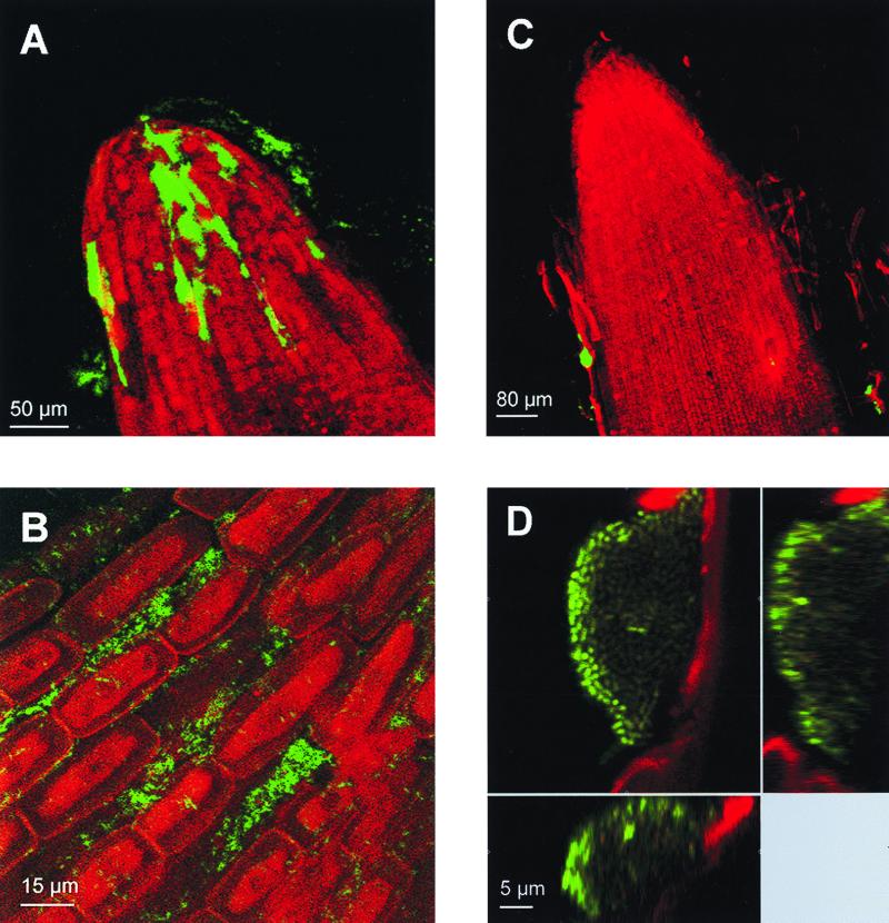
FIG. 4.
In situ visualization of active P. putida cells on the root surfaces of barley seedlings: visualization of Gfp-tagged P. putida CRR300 (PA1/04/03::gfpmut3*) (A and B) and P. putida SM1700 (rrnBP1::gfp[AGA]) (C and D) cells colonizing the root tips of 2-day-old barley seedlings. The green fluorescence emitted by the cells and the red autofluorescence emitted by the root material were visualized by SCLM. (A and C) xyz scan pictures (magnification, ×200) of root tips colonized by P. putida CRR300 and SM1700, respectively. (B and D) xyz pictures (magnification, ×630) of bacterial cells on the root surfaces. Vertical sections of the colony shown in panel D are shown to the right and below.

