Figure 1.
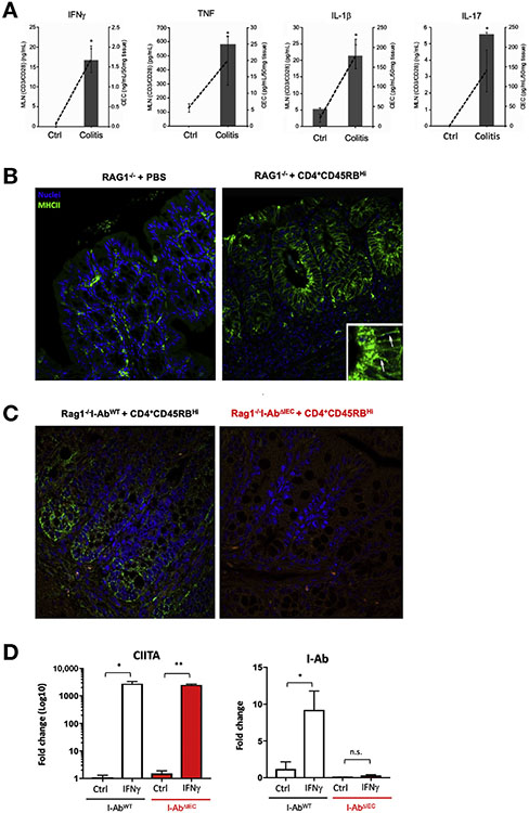
Expression of MHCII in the colonic epithelium in adoptive T-cell transfer colitis. (A) Secretion of IFNγ, TNFα, IL1β, and IL17 by the CD3/CD28-stimulated MLN cells (bars, left axis) and by the colonic explants (dashed line, right axis). (B) Representative immunofluorescence imaging (n = 4). Arrows in the magnified inset show green MHCII signal. (C and D) MHCII ablation in I-AbΔIEC mice. (C) Representative immunofluorescence of colonic sections (n = 8) showing complete loss of epithelial MHCII expression in adoptively transferred Rag1−/−I-AbΔIEC mice (right) as compared with Rag1−/−I-AbWT mice (left). (D) qRT-PCR analysis of CIITA and I-Ab mRNA expression in control and IFNγ-stimulated (100 U/mL for 18 hours) colonoids prepared from I-AbWT and I-AbΔIEC mice. TATA-box binding protein (TBP) was used as housekeeping gene. Error bars indicate standard deviation. *P < .05, **P < .01, Student t test.

