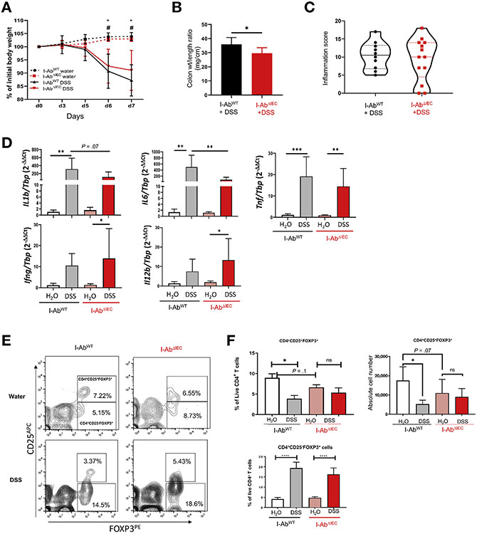
Figure 2.
Deletion of MHCII in IEC offers modest protection in acute DSS colitis. I-AbWT and I-AbΔIEC mice were treated with 3% DSS in drinking water for 7 days. Data representative of 2 independent experiments (n = 10–13 mice in DSS group and n = 11–17 mice in H2O group). (A) Weight loss expressed as percentage relative to the initial weight (*P < .005 or 0.001 H2O vs DSS-treated I-AbWT mice on days 6 and 7, respectively; #P < .005 H2O vs DSS-treated I-AbΔIEC mice on days 6 and 7. (B) Colon length/weight ratio in DSS-treated mice. (C) Violin plot of the colonic inflammation score (solid lines indicate medians; dashed lines indicate quartiles). Slides were scored blindly, and total scores calculated as the sum of proximal and distal colon score. (D) qRT-PCR analysis of colonic IL1β, IL6, TNFα, IFNγ, and IL12b mRNA in I-AbWT and I-AbΔIEC mice. Results expressed as relative expression normalized to TATA-box binding protein (TBP) mRNA. *P < .05, **P < .01, ****P < .0001 (1-way analysis of variance [ANOVA] followed by Bonferroni multiple comparison test). (E) Representative flow cytograms demonstrating the CD4+CD25+Foxp3+ cells and CD4+CD25−Foxp3+ regulatory T cells in the cLP of I-AbWT and I-AbΔIEC mice. (F) Summary analysis of CD4+CD25+Foxp3+ and CD4+CD25−Foxp3+ cells in I-AbWT and I-AbΔIEC on water or DSS. *P < .05, ****P < .0001 (1-way ANOVA followed by Bonferroni multiple comparison test).

