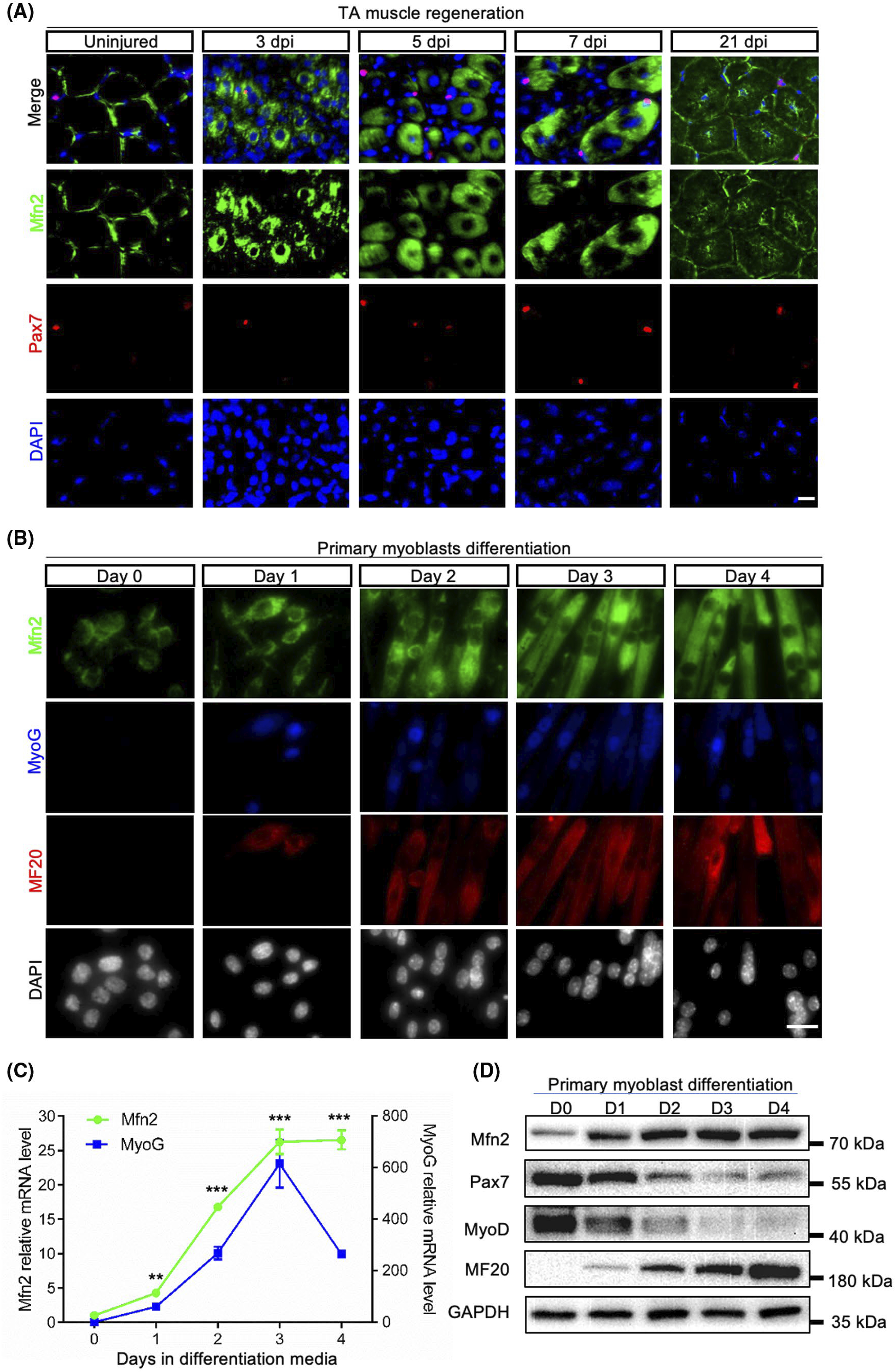
FIGURE 1.

Mfn2 expression is upregulated during muscle regeneration and myoblast differentiation. A, Mfn2 immunofluorescence in of uninjured and injured TA muscle sections at 3, 5, 7, and 21 days post-injury (dpi). Scale bar: 20 μm. B, Mfn2 immunofluorescence in primary myoblasts in growth medium (Day 0) and after differentiated for 1–4 days. Scale bar: 20 μm. C, qRT-PCR analysis of relative mRNA levels of Mfn2 and Myog genes (n = 3 each group). Data are shown as mean ± SEM (t test: **P < .01, ***P < .001). D, Western blots showing relative levels of Mfn2 and myogenesis-related proteins (Pax7, MyoD, and MF20) at various stages of myoblast differentiation
