FIGURE 4.
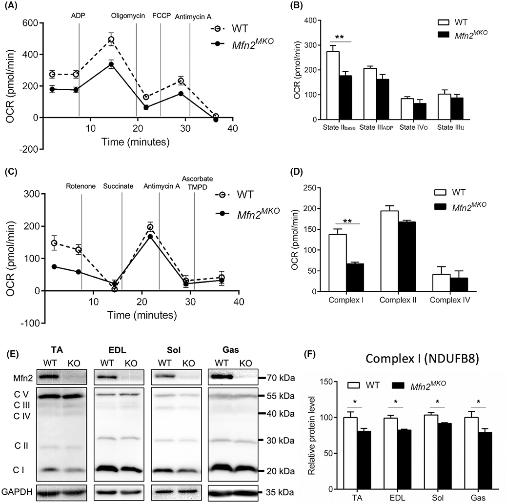
Loss of Mfn2 reduces the level of mitochondrial complex I protein and affects electron transport. A, Seahorse coupling assay showing oxygen consumption rates (OCR) in mitochondria isolated from WT and Mfn2MKO Sol muscles. B, Quantification of OCR in state IIbase (Basal respiration), state IIIADP (ADP-stimulated respiration), state IVo (non-ADP-stimulated respiration, after oligomycin treatment), and state IIIu (uncoupled respiration, after FCCP treatment) (n = 3 pairs of mice). C, Seahorse electron transport chain assay showing oxygen consumption rates (OCR) in mitochondria isolated from WT and Mfn2MKO Sol muscles. D, Quantification of OCR in complex I, complex II, and complex IV (n = 3–5 pairs of mice). E, Western blots showing the protein levels of complexes I (NDUFB8), II (SDHB) III (UQCRC2), IV (MTCO1) and V (ATP5A) in TA, EDL, Sol and Gas muscles of WT and Mfn2MKO mice. F, Quantification of the relative levels of complex I (normalized to GAPDH) in TA, EDL, Sol, and Gas muscle (n = 4–6 pairs of mice). All data are shown as mean ± SEM (t test: *P < .05, **P < .01)

