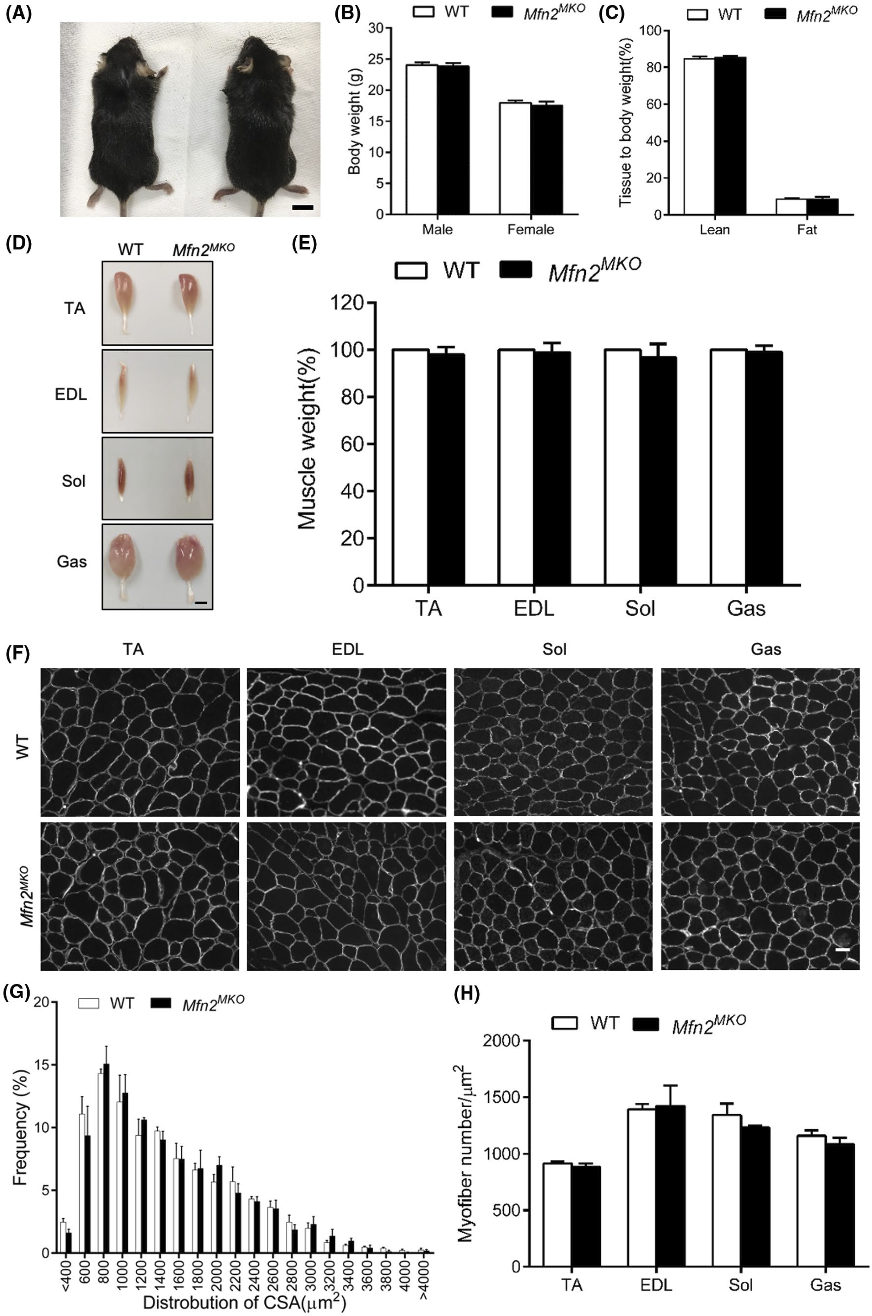
FIGURE 6.

Conditional knockout of Mfn2 in myogenic progenitors does not affect muscle development. A, Representative images of WT and Mfn2MKO mice, scale bar: 1 cm. B, Body weights of male and female WT and Mfn2MKO mice at 2 months old (n = 10 pairs of mice). C, EchoMRI analysis showing ratios of lean and fat mass relative to body weight (n = 3 pairs of mice). D and E, Representative images (D) of TA, EDL Sol, and Gas muscles isolated from adult WT and Mfn2MKO mice (scale bar: 2 mm) and their relative weights (WT weights are normalized to 100, n = 6–8 pairs of mice). F, Dystrophin immunofluorescence outlining myofiber membrane to reveal myofiber size in WT and Mfn2MKO muscles. Scale bar: 50 μm. G, Distribution of myofiber cross-sectional areas (CSA, μm2) of Sol muscles (n = 4 each group). H, Average numbers of myofiber per μm2 in various muscles (n = 5–6 pairs of mice). All data represent mean ± SEM
