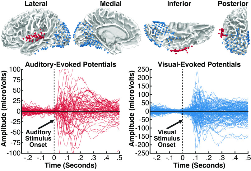
Figure 8.
Experiment 2. Top: intracranial electrodes from 13 patients, displayed on an average brain (all electrodes projected into the left hemisphere). Each colored sphere reflects a single electrode contact included in analyses, localized in auditory areas (red, 73 electrodes) or visual areas (blue, 151 electrodes). Auditory electrodes were limited to those located proximal to the superior temporal gyrus or neighboring white matter and showing a significant event-related potential (ERP) to sounds beginning at less than 120 ms. Visual electrodes were limited to those located in occipital, parietal, or inferior temporal areas and showing a significant ERP to visual stimuli beginning at less than 120 ms. Bottom: ERP responses at all auditory electrodes (red) in evoked during a passive listening task, and at all visual electrodes (blue) evoked during a visual task; the time zero indicates stimulus onset in each task.

