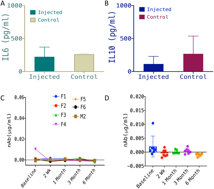
Fig. 6: No detectable inflammatory response in vitreous humor or immune response in plasma of vMCO1 injected mice.
Quantitative comparison of IL-6 (pro-inflammatory marker) (A) and IL-10 (anti-inflammatory marker) (B) in vitreous humor of rd10 mice injected with vMCO1 (1.0 x 10⁹ vg in eye) and non-injected control, 6 months after vMCO1 injection (at the age of 12 weeks). Average ± SD. N=7. (C) Longitudinal monitoring of neutralizing Antibody (nAb) level in serum of rd10 mice injected with vMCO1 (1.0 x 10⁹ vg in eye) before and after injection (F: Female; M: Male) at 12 weeks of age. (D) Scatter plot showing the mean and variation of measured neutralizing antibody concentration in serum of vMCO-010 injected rd10 mice at each time point. N=7.

