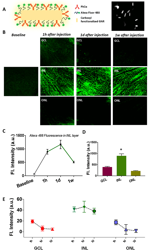
Fig. 2. Kinetics of retinal layer-specific fGNR binding shows peak after 1d of injection, which declines after 1 wk.

(A) Schematic of fGNR functionalized with PKCα antibody for bipolar cell specific binding along with SEM image of fGNRs. (B) Layer-specific binding of fGNRs after 1 hour, 1 day and 1 week of intravitreal injection in mice. Z-section of confocal microscopy imaging reveals the highest presence of GNR in the inner nuclear layer (except in baseline) compared to other retinal layers (such as ganglion cell layer and photoreceptor layer). (C) Change in layer-specific fluorescence (fluorescent tagged fGNRs) over time in INL layer (baseline fluorescence of the INL layer is before injection of GNRs). (D) Layer-specific binding of fGNRs after 1d of injection. (E) Longitudinal monitoring of functionalized GNRs in different retinal layer. N=3 animals. *p<0.05.
