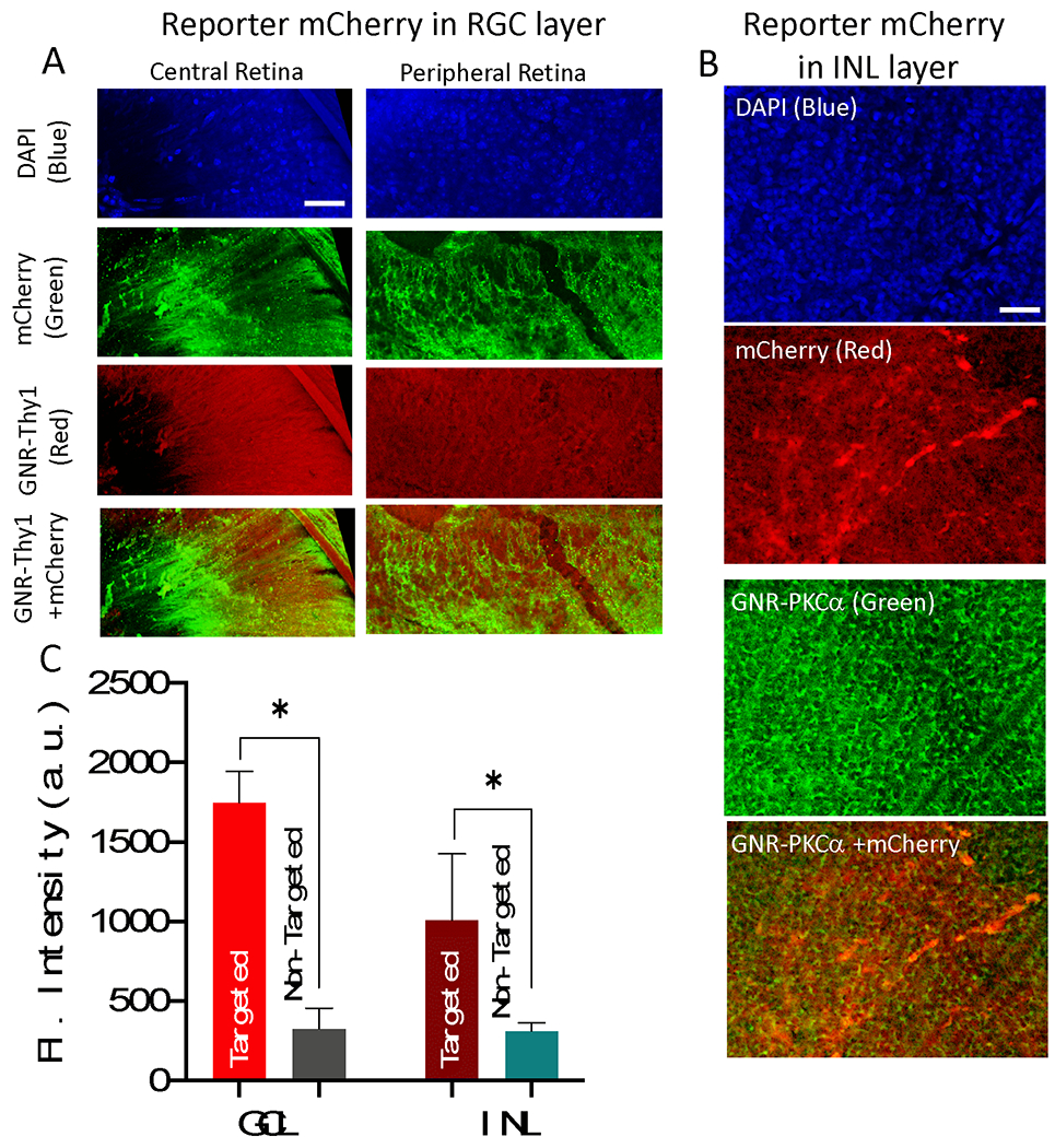
Fig. 3. Triple selective optical delivery via (i) cell-specific functionalization of gold nanorods, (ii) Spatially localized laser irradiation, and (iii) delivery laser wavelength tuned to SPR of GNRs.

(A) RGC specific Thy1 antibody functionalized GNRs (labeled with Alexa 555-Red), MCO expression achieved by 800 nm CW laser (30 mW, 60 s) visualized by mCherry antibody staining (Green), 1 week after gene delivery. (B) Bipolar cell specific PKCα antibody functionalized GNRs (labeled with Alexa 488-Green), ON-bipolar cell specific MCO expression achieved by 850 nm CW laser (total dose =1800 mJ/mm2: 30 mW for 60 sec, 1 mm2 area) visualized by antibody staining (Red), 1 week after gene delivery. Scale bar: 50 μm. (C) The quantitative comparison of MCO (mCherry reporter) expression in RGC layer and INL versus non-targeted area. N=3 animals. *p<0.05
