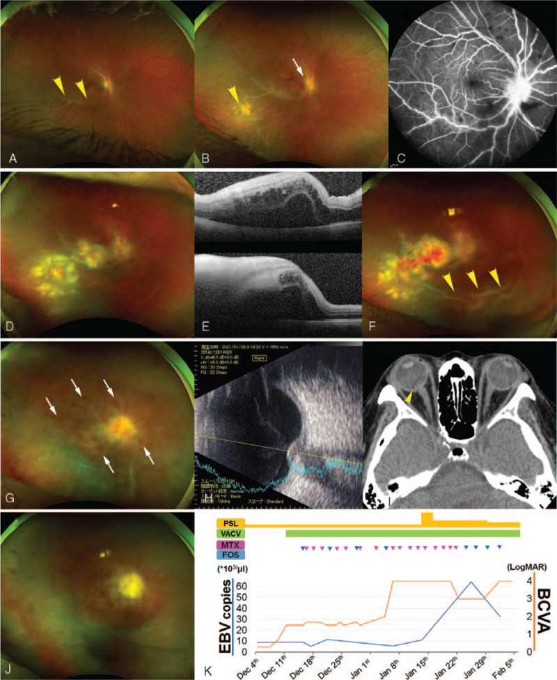
Figure 1.
Clinical course of the patient with Epstein–Barr virus intraocular infection. A unilateral exudative change along the vessel appears at the temporal retina of the 44-year-old patient on October 30, 2020 (A, arrowheads). A snowball-like vitreous opacity, recent retinal spot (B, arrowhead), and peripapillary exudative changes (B, arrow) have developed by November 26, 2020 (B), detected on fluorescein angiography (C). By December 11, 2020, the retinal lesions, such as retinal infiltration and hemorrhage, have progressed to involve the macula (D), with severe edema and exudative detachment observed on optical coherence tomography (E, upper, horizontal image; lower, vertical image). Retinal hemorrhage and vitreous opacity have been observed on December 14, 2020 (F, arrowheads). On January 6, 2021, she has developed central retinal vein occlusion (G, hemorrhage is pointed by arrows) related to the swelling of the optic-nerve papillae recorded, using echography (H) and computed tomography (I, arrowhead). However, the vitreous infiltration is suppressed (G). The activity of the optic-nerve lesion and the retinitis and vitreous infiltration are suppressed on January 28, 2021 (J). (K) depicts the EBV copy numbers in the aqueous humor and best-corrected visual acuity over time, in relation to the administered drugs. Each triangle denotes the intravitreal injection (MTX, pink; foscarnet, blue). EBV = Epstein–Barr virus, FOS = foscarnet, MTX = methotrexate, VACV = valaciclovir hydrochloride.

