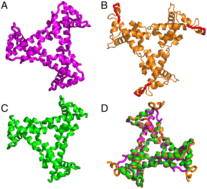
Fig. 1.
X-ray structure of HIV-1 myrMA112. Three independent monomeric chains (a, b, and c) of MA112 were found in the asymmetric unit of the crystal. Trimeric assemblies are shown of chain a (A), chain b (B), and chain c (C). These assembled units are generated by imposing crystallographic symmetry. In B, the complete N terminus of myrMA112 is shown with attached myr group (red) and linkage to the N-terminal glycine shown in stick model. For chain b, well-defined electron density was observed for the entire N terminus (SI Appendix, Fig. S1). For chains a and c, electron density for the myr group as well as residues 2–5 and 2–9, respectively, was not observed presumably due to flexibility of the N terminus. (D) Overlay of the three trimers showing that the structures of the MA molecules and the trimer arrangements are nearly identical.

