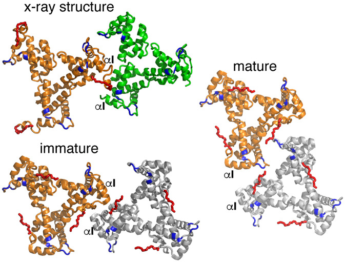
Fig. 6.
Comparison of trimer–trimer contacts in the lattice. Cartoon illustration of the HIV-1 myrMA trimer–trimer units obtained by the X-ray structure and those reconstructed from the cryotomography data of the immature (PDB ID code 7OVQ) and mature (PDB ID code 7OVR) particles. The orange trimers are in an identical orientation. As shown, the trimer–trimer relationship and interface in the X-ray structure is relatively similar to that of the immature particle. The HBR and myr groups are shown in blue and red, respectively.

