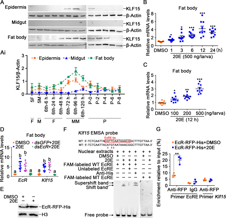
Fig 1. 20E upregulated the expression of Klf15.
A. The protein profiles of KLF15 in the epidermis, midgut, and fat body detected using western blotting after 12.5% SDS-PAGE. 5F: fifth instar feeding larvae; 5M: fifth instar molting larvae; 6th-6 h to 120 h: sixth instar larvae at different stages; P0 to P8: 0 to 8-day-old pupae. F: feeding; M: molting; MM: metamorphic molting; P: pupae. Ai. Quantification of KLF15 in A using Image J software. B. Time course of the Klf15 expression in the fat body after 20E (500 ng/larva) induction. DMSO was used as the control. C. The expression of Klf15 in the fat body under stimulation with different concentrations of 20E for 12 h. D. Knockdown of EcR in the fat body by dsEcR (3 μg/larva) followed by stimulation with 20E (500 ng/larva) for 12 h to detect the expression of Klf15. E. Nuclear proteins from EcR-RFP-His overexpressed cells were extracted for EMSA. F. EcRE on the Klf15 promoter bound to EcR detected by EMSA assay. WT and MT represent EcRE probe and EcRE mutant probe, respectively. G. ChIP assay showing 20E promoted Klf15 expression via EcR binding to EcRE and detected by qRT-PCR. Primer EcRE is the sequence containing EcRE. Primer Klf15, as non-EcRE control targeting to Klf15 open reading frame (ORF).

