. 2022 May 26;11:e74687. doi: 10.7554/eLife.74687

Figure 2. HAESA-LIKE 3 (HSL3) forms a CTNIP-induced receptor complex with BAK1.

(a) Schematic representation of BAK1-GFP immunoprecipitation in the (1) absence or (2) presence of CTNIP4 treatment to identify protein associations induced by CTNIP. Figure generated using Biorender. (b) HSL3-specific spectral counts identified in four independent biological replicates where BAK1-GFP was pulled down in the presence or absence of 1 μM CTNIP4 treatment. Circle diameter is proportional to the number of replicates. Red lines indicate the mean spectral counts for each treatment. p-Values indicate significance relative to the untreated control in a two-tailed t-test. (c) Affinity purification of BAK1 with HSL3-GFP from HSL3-GFP seedlings treated with 1 μM CTNIP448-70 or water for 10 min. Western blots were probed with antibodies α-GFP and α-BAK1. This experiment was repeated three times with similar results. (d) Isothermal titration calorimetry (ITC) summary table of HSL3 vs. CTNP448-70, CTNP4C58S/C68S and INFLORESCENCE DEFICIENT IN ABSCISSION (IDA) peptides, and contribution of the BAK1 co-receptor to the ternary complex formation. Kd, (dissociation constant) indicates the binding affinity between the two molecules considered (nM). The N indicates the reaction stoichiometry (N=1 for a 1:1 interaction). The values indicated in the table are the mean ± SEM of two independent experiments. (e) ITC experiments of HSL3 vs. CTNIP4 and CTNIP4C58S/C68S, in the absence and presence of the co-receptor BAK1. GFP, green fluorescent protein.

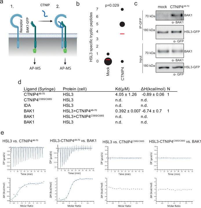
Figure 2—source data 1. HSL3-specific spectral counts.
Figure 2—source data 2. Westernblots showing affinity purification of BAK1 with HSL3-GFP.

Figure 2.

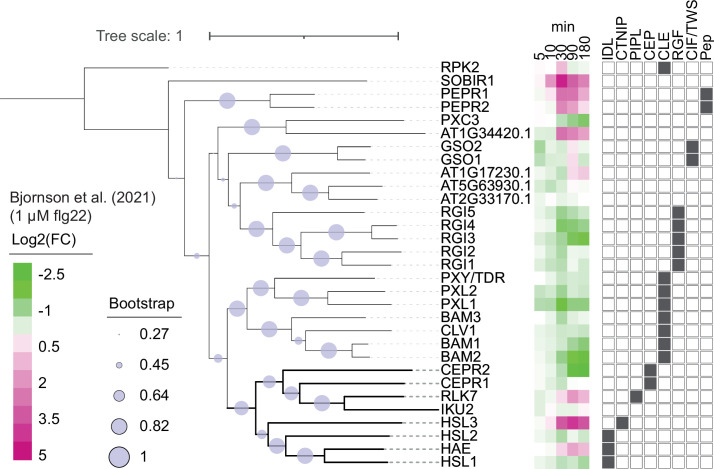
Figure 2—figure supplement 1. Phylogeny of Arabidopsis leucine-rich repeat receptor kinase (LRR-RK) subfamily XI.

Figure 2—figure supplement 1.

Phylogeny of full-length protein sequences of the Arabidopsis LRR-RK subfamily XI. Sequences were aligned using MUSCLE and a phylogeny was inferred using the maximum-likelihood method and JTT matrix-based model conducted in MEGAX. 1000 bootstraps were performed and are indicated based on the size of the blue circles. Expression of these receptors in response to 1 μM flg22 treatment was extracted from Bjornson et al., 2021, and is represented in a heat map. Known ligands for LRR-RK subfamily XI are highlighted to the right (Butenko et al., 2003; Cho et al., 2008; Crook et al., 2020; Doblas et al., 2017; Doll et al., 2020; Hou et al., 2014; Krol et al., 2010; Morita et al., 2016; Mou et al., 2017; Nakayama et al., 2017; Ogawa et al., 2008; Okuda et al., 2020; Ou et al., 2016; Qian et al., 2018; Rojo et al., 2002; Roman et al., 2022; Santiago et al., 2016; Shinohara et al., 2016; Song et al., 2016; Tabata et al., 2014; Yamaguchi et al., 2010; Yamaguchi et al., 2006; Zhang et al., 2016).
Figure 2—figure supplement 1—source data 1. Flg22-induced expression data of LRR-RK subfamily XI.

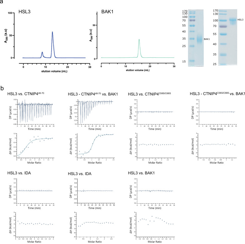
Figure 2—figure supplement 2. Isothermal titration calorimetry (ITC) independent experiments and purification of HAESA-LIKE 3 (HSL3) and BAK1 used in the binding experiments.

Figure 2—figure supplement 2.

(a) Analytical size exclusion chromatography (SEC) of the ectodomains of HSL3 and BAK1. An SDS-PAGE of the two proteins is shown alongside. (b) ITC raw thermograms of experiments shown in the ITC table summary in Figure 2d.

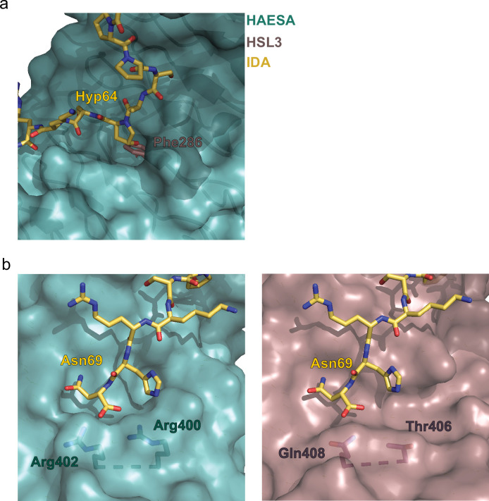
Figure 2—figure supplement 3. Structural comparison of the binding pockets between the receptors HAESA and HAESA-LIKE 3 (HSL3).

Figure 2—figure supplement 3.

(a) The hydroxyproline pocket required for anchoring the INFLORESCENCE DEFICIENT IN ABSCISSION (IDA) peptide to the HAESA receptor is missing in HSL3. Close view of the binding pocket of the structural superimposition of the HAESA-IDA complex (PDB:5IXQ) and a homology model of HSL3 (AlphaFold: https://alphafold.ebi.ac.uk/). The HAESA receptor is depicted in surface representation in teal blue, IDA in yellow sticks, and HSL3 in magenta cartoon. In HSL3, the hydroxyproline pocket is replaced by the bulky residue Phe286, colliding with the potential anchoring of the IDA peptide to the receptor. (b) The conserved RxR motif necessary for the coordination of the COO- group the last Asn in IDA is not present in the HSL3 receptor. Zoom in of the C-terminal region of the peptide-binding surface of HAESA (teal blue) (left panel) and HSL3 (magenta) (right panel). In HAESA the motif RxR closes the binding pocket allowing for the coordination of the C-terminal of IDA. In HSL3 this structural motif is substituted by the residues Thr406 and Gln408, leaving the binding surface open to potentially accommodate a longer peptide ligand. Figures were done using the PyMOL Molecular Graphics System, Version 2.0 Schrödinger, LLC.