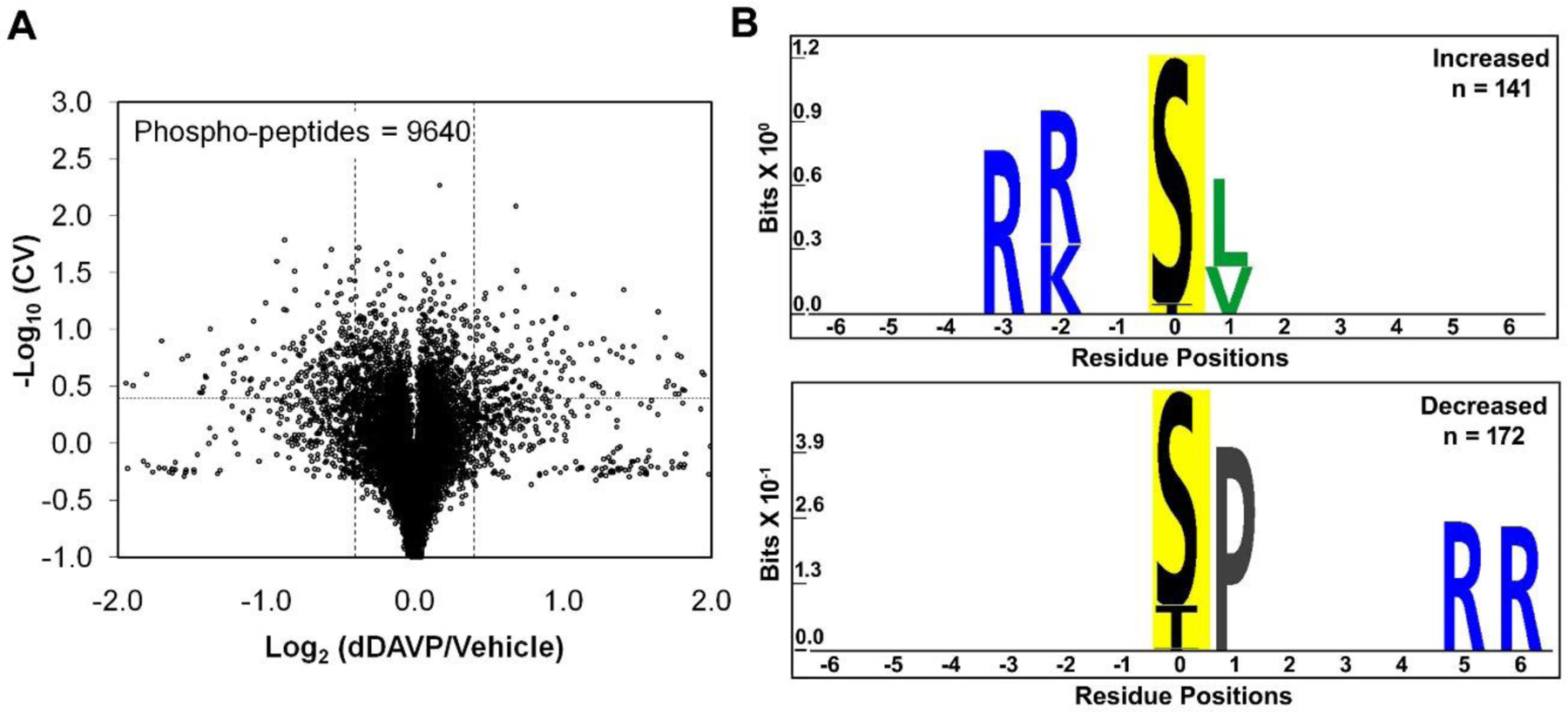
Figure 2.

Vasopressin effects on phosphoproteome of mpkCCD cells. A. Torch plot for phosphopeptides quantified across all three pairs (dDAVP vs. vehicle) of mpkCCD cell clones. Vertical dashed lines show log2(dDAVP/Vehicle) of 0.4, and −0.4, while the horizontal line represents −log10(CV) of 0.4 (CV=0.4). These phosphopeptides are found in 295 distinct phosphoproteins. Out of 9640 quantified phosphopeptides, 429 were altered (increased, 187; decreased, 242) according to the thresholds defined above. These phosphopeptides consisted of 332 mono-phosphopeptides (increased, 153; decreased, 179) and 97 multi-phosphorylated peptides. B. Motif analysis shows the amino acid residues over-represented in sets of unique mono-phosphopeptides whose abundances are significantly increased (141 phospho-sites, corresponds to 153 mono-phosphopeptides; upper panel) and decreased (172 phospho-sites, corresponds to 179 mono-phosphopeptides; lower panel). PTM-Logo was used with whole mouse dataset as background and Chi-square cutoff was set at 0.0001.
