Figure 5.

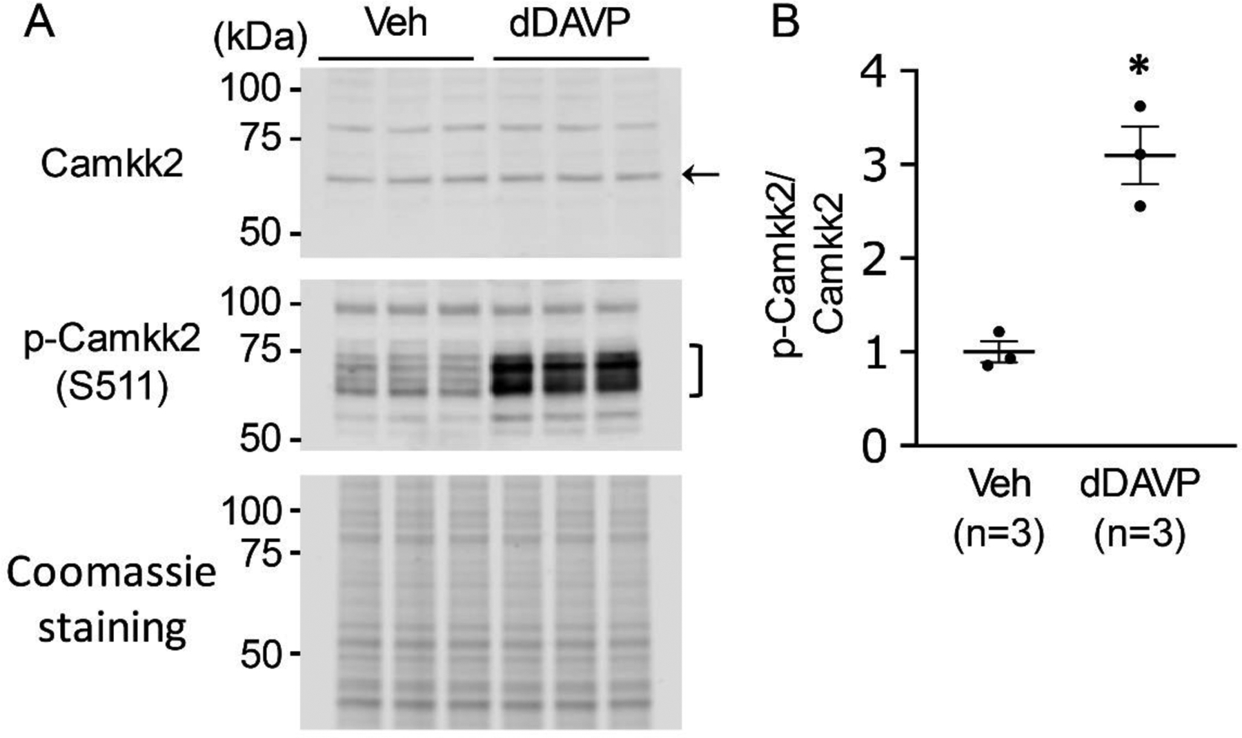
Immunoblot analysis to detect dDAVP-induced phosphorylation on Camkk2. A. Camkk2 and pS511-Camkk2 immunoblot from dDAVP- and vehicle-treated mpkCCD cells. The gels were stained with Coomassie stain to check equal loading among different samples. B. Separated scatter plot showing the ratio of normalized immuno-reactivities (mean ± SEM in arbitrary units) of pS511-Camkk2 and Camkk2. Unpaired t-test was used to determine significant difference between dDAVP and vehicle-treated samples. dDAVP caused a significant increase in phosphorylation of S511 of Camkk2 in mpkCCD cells. n, experimental replicate for vasopressin stimulation experiment. *P < .05. A number of immunoblots using phospho-specific antibodies have been reported previously (https://esbl.nhlbi.nih.gov/ESBL/WB-data/).
