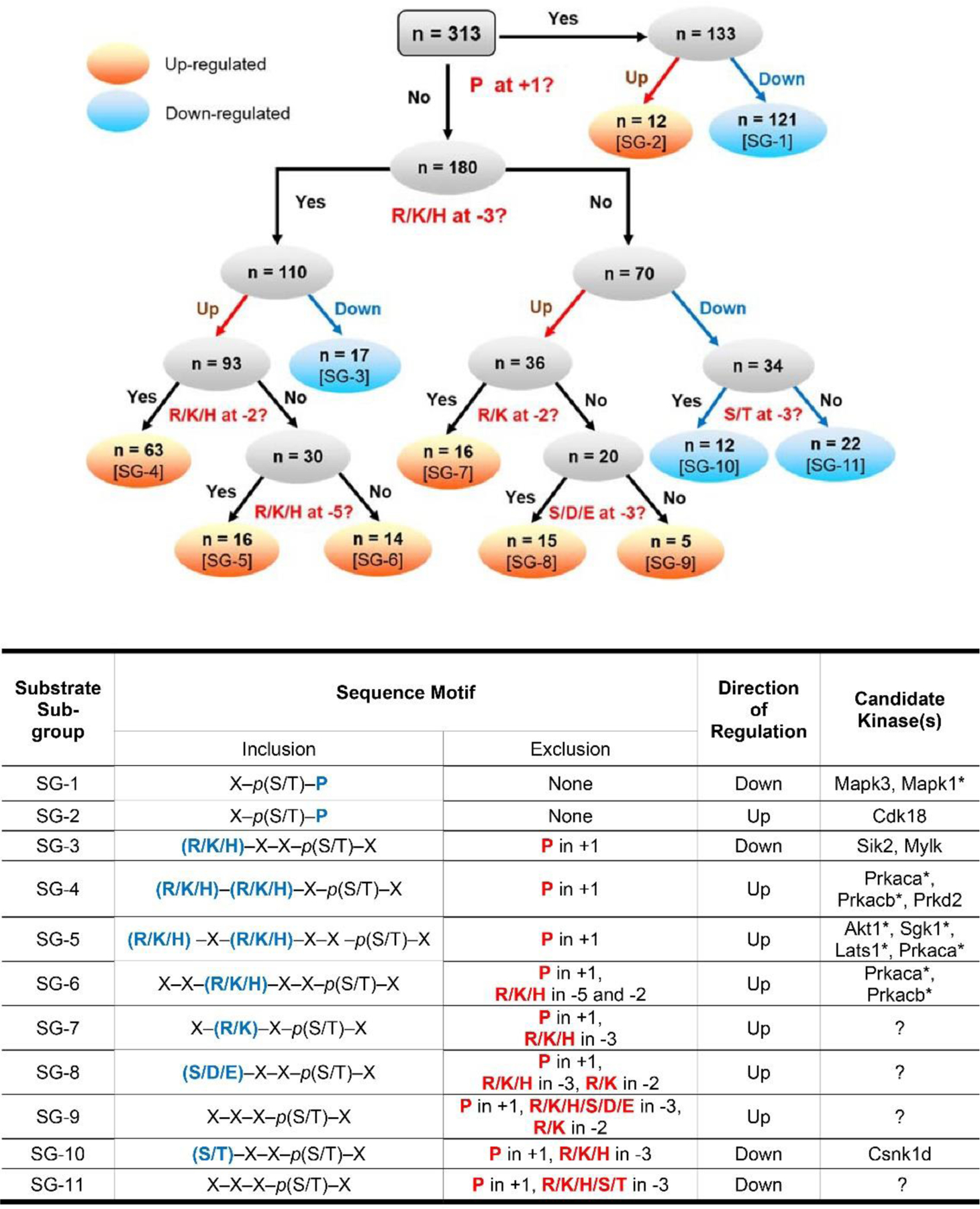
Figure 7.

Rule-based classification of vasopressin-regulated phosphorylation sites. Top panel: All 313 regulated single phosphorylation sites were classified into 11 sub-groups (numbered SG-1 to SG-11 in square brackets). This classification is based on the direction of change (up-regulated or down-regulated) and sequence surrounding the phosphorylated S or T in position +1, −2, −3 and −5. Seven subgroups contain up-regulated phospho-sites (orange) while 4 sub-groups contain down-regulated phospho-sites (blue). Bottom panel: The table presents the sequence motifs in the ‘inclusion’ column (amino acids in blue color) for all 11 sub-groups of substrates along with the candidate kinases. Amino acid residues that are excluded from specific positions are indicated in the exclusion column. *Kinases that are not listed in Table 1.’?’, no known kinase.
