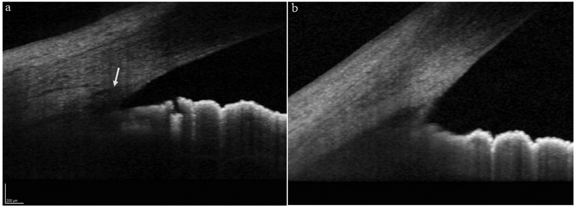
Figure 2 –

High-definition anterior segment OCT images showing a) normal appearing angle with the white arrow pointing towards the Schlemm’s canal, b) an angle of a JOAG patient showing abnormal thickness in the region of trabecular meshwork and without a visible Schlemm’s canal.
