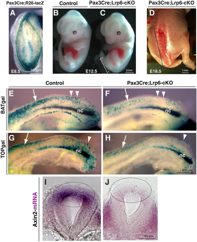
Fig. 1.

Spinal bifida aperta and diminished canonical Wnt signaling by conditional ablation of Lrp6 in Pax3-expressing dorsal neural folds. (A) Dorsal–posterior view of an X-gal-stained (blue) E8.5 embryo for genetic fate mapping of Pax3Cre/+;Rosa26-lacZ demonstrates the Cre recombination pattern in the dorsal region of the recently closed and pending-closing posterior neuropore (PNP; indicated by dashed line). (B-D) The conditional mutants of Pax3Cre/+;Lrp6-cKO embryos exhibit open spinal neural tube defects (NTDs), as shown at E12.5 and E18.5. Dashed line brackets indicate the open lesion regions. (E-H) Sagittal caudal bodies of X-gal-stained Wnt/β-catenin signaling reporters BATgal or TOPgal show higher activities in the littermate control embryos (E,G) and diminished activities in the Pax3Cre/+;Lrp6-cKO embryos (F,H) at E9.5. Arrows indicate recently closed dorsal neural tube regions. Arrowheads indicate the closing or pending-closing regions. nc, notochord. (I,J) Transverse sections show in situ hybridization signal of a Wnt/β-catenin target and feedback gene Axin2, which is high in the dorsal PNP of a littermate control (dashed line oval in I) and low in the mutant PNP (dashed line oval in J) at E9.5.
