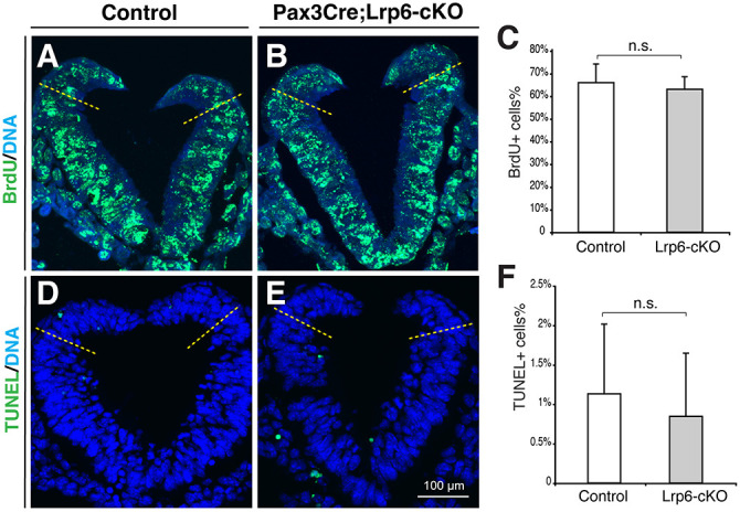
Fig. 6.

Proliferation and apoptosis at the PNP closure sites of the littermate controls and Pax3-Cre;Lrp6-cKOs at E9.5. (A-C) BrdU incorporation and detection experiments show no significant differences in proliferating cells in the dorsal PNPs above the dorsolateral hinge points (dashed lines in A,B, transverse PNP sections) between the control and mutant embryos. (D-F) TUNEL assays demonstrate no significant differences in apoptotic cells (green in D,E) in the dorsal PNPs between the control and mutant embryos. n.s., no statistical significance (P>0.05; unpaired, two-tailed Student's t-test).
