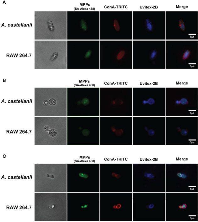
Figure 5.
Mannose purified proteins (MPPs) of Acanthamoeba castellanii and RAW 246.7 macrophages co-localized with a mannose-specific lectin Concanavalin A. Fluorescence microscopy images demonstrate the cell wall of the fungi (A) Candida albicans; (B) Cryptococcus neoformans and (C) Histoplasma capsulatum stained in blue (Uvitex 2B); MPPs of phagocytes stained in green upon incubations with yeasts, followed by the streptavidin-Alexa 488 conjugate; and the mannosylated components of the yeast cell wall stained in red, after incubations with ConA-TRITC. Scale bar= 5μm.

