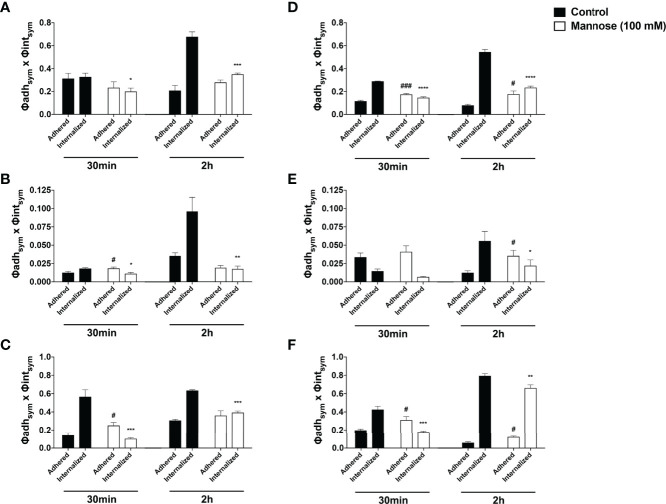
Figure 8.
The mannose receptors participates on fungal internalization by Acanthamoeba castellanii and macrophages Phagocytosis assays were performed with either (A–C) A. castellanii or (D–F) RAW 264.7 upon addition of NHS-Rhodamine labeled (A, D) Candida albicans, (B, E) Cryptococcus neoformans and (C, F) Histoplasma capsulatum, in the absence (Control group, black bars) and presence of 100 mM soluble mannose (Mannose 100 mM, white bars) and analyzed after 30 min, to evaluate the initial fungal adhesion, and 2h, to evaluate the late interaction. Symmetrized adhesion and phagocytic indices were determined and the mannose inhibition groups were compared to controls. The statistical significances are represented above each group, where *p < 0.05, **p < 0.01, ***p < 0.001 or ****p < 0.0001 indicate statistically significant decrease upon mannose inhibition; #p < 0.05, ###p < 0.001 indicate statistically significant increase upon mannose inhibition; ns = not significant). The bars represent the average of three independent experiments, performed in triplicates.

