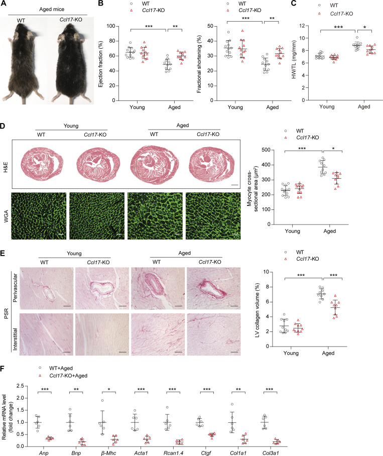
Figure 2.
Ccl17-KO mice were protected from age-related cardiac hypertrophy and HF. (A) Representative images of aged (21-mo-old) C57BL/6J background WT and Ccl17-KO mice. (B) Measurement of EF and FS in C57BL/6J background WT and Ccl17-KO mice at 4 mo (young) or 21 mo (aged; n = 11–12 biological replicates). (C) HW/TL ratios of C57BL/6J background WT and Ccl17-KO mice at 4 mo (young) or 21 mo (aged; n = 11–12 biological replicates). (D) Histological analysis of heart sections of C57BL/6J background WT and Ccl17-KO mice at 4 mo (young) or 21 mo (aged). Heart cross-sections were stained with H&E (top) to analyze hypertrophic growth of hearts, scale bars: 1 mm. WGA (bottom) staining of heart sections and corresponding group quantification data (right) to determine cell boundaries (n = 10–12 biological replicates). Scale bars: 30 µm. (E) PSR (left) staining and corresponding group quantification data (right) of C57BL/6J background WT and Ccl17-KO mice at 4 mo (young) or 21 mo (aged; n = 9–10 biological replicates). Scale bars: 50 µm. (F) qRT-PCR was performed to analyze the mRNA levels of cardiac function (Anp, Bnp), hypertrophy (β-Mhc, Acta1, Rcan1.4), and fibrosis (Ctgf, Col1a1, Col3a1) markers in aged (21-mo-old) C57BL/6J background WT and Ccl17-KO mice (n = 6 biological replicates). All data are presented as the mean ± SD. Statistical significance was tested using one-way ANOVA, followed by the Bonferroni post hoc test (B–E). The unpaired Student’s t-test was used for equal variance and the Welch t-test for unequal variance (F). *, P < 0.05; **, P < 0.01; ***, P < 0.001.

