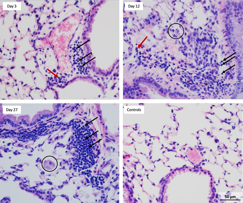
Fig. 7.
Micrographs of lung sections stained with H&E. Development of progressive perivascular aggregates (black arrows), mainly lymphocytes, indicating chronic inflammation was observed from Day 3 – 27. Occasional neutrophils (red arrows) were seen mainly at earlier time points (Day 3 and Day 12) indicating an acute inflammation. Control mice showed no lesions with clear alveolar spaces and no overt recruitment of lymphocytes or neutrophils. Similarly, as seen in BAL fluid, macrophages became more enlarged and activated with foamy vacuoles (black circles). All images were taken at the same magnification

