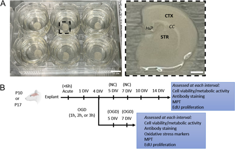
Fig. 1.
Schematic of OWH slice culture methodology and experimental workflow. A Left: Coronal slices containing cortex (CTX) and striatum (STR) are plated onto PTFE membrane inserts. Right: a single OWH brain slice after explantation. Although not imaged in this study, hippocampal (HIP) and corpus callosum (CC) regions are also labeled for reference. Scale bars = 15 cm and 5 cm. B Experimental workflow. Slices from postnatal (P) day 10 or P17 aged rat donors are cultured up to 14 days, performing OGD on 4 DIV with endpoints at 5 days in vitro (DIV) (24 h post-OGD), 7 DIV (72 h post-OGD), and 10 DIV

