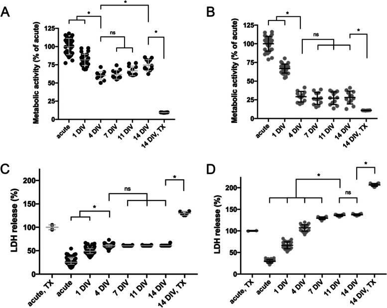
Fig. 2.
Metabolic activity and LDH release profiles for P10 and P17 brain slices for 14 DIV following explantation. Metabolic activity values are normalized to the metabolic activity at the acute timepoint. LDH release (%) values are normalized to the LDH release of acute slices immediately treated with TX-100. A-B Metabolic activity of (A) P10 and (B) P17 slices. Error bars represent the mean ± SD. C-D % LDH release profile of (C) P10 and (D) P17 slices. Error bars represent median ± IQR (C) and mean ± SD (D). n = 6–42 OWH slices. * denotes significant differences (p < 0.05). For metabolic activity data (A&B) and LDH release data in P10 brain slices (C), comparisons were made between the 4DIV timepoint and all other time points. For LDH release data in P17 brain slices (D), comparisons were made between the 11DIV timepoint and all other time points. In all instances (A-D), comparisons were also made between the 14DIV timepoint and the 14DIV, TX-100 treated group

