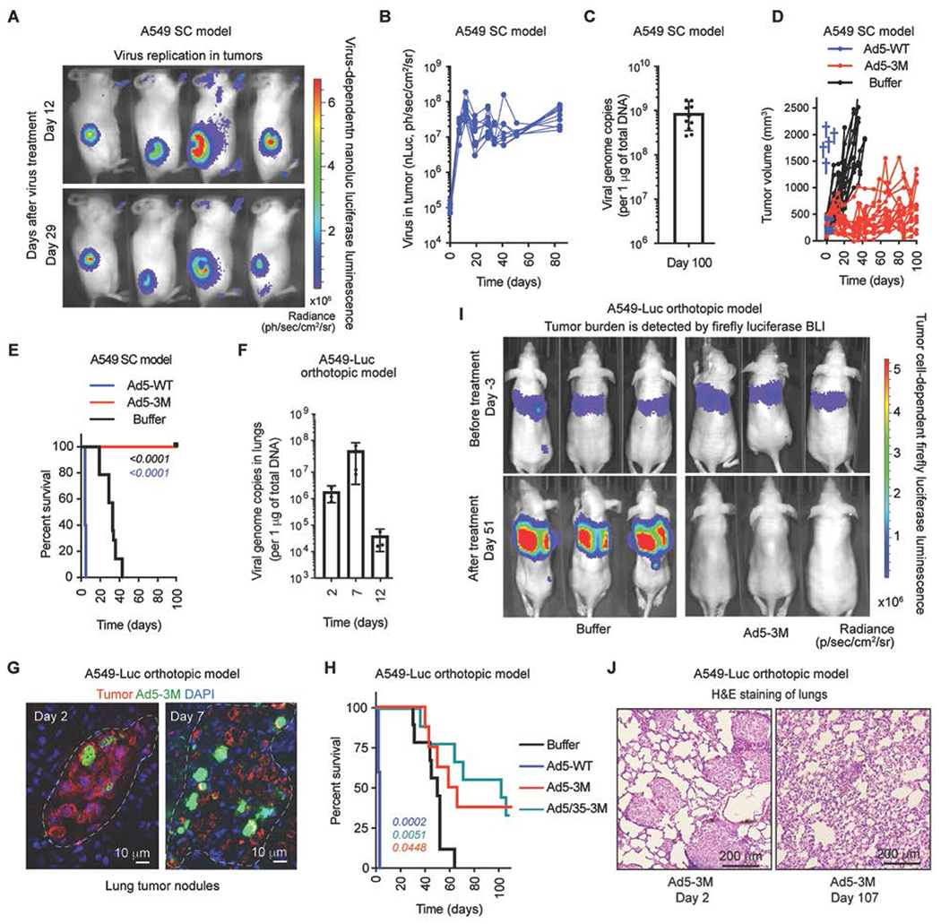
Fig. 5. Ad5-3M transduces tumors, suppresses tumor growth, and extends survival of mice with localized and disseminated tumors after systemic delivery.

(A) In vivo bioluminescence imaging (BLI) of subcutaneous tumor-bearing mice at days 12 and 29 post Ad5-3M treatment. The color indicates bioluminescence intensity of virus-encoded nano-luciferase. SC – subcutaneous. (B) Activity of virus-encoded nano-luciferase in tumor-bearing mice treated with Ad5-3M (n = 10). (C) The amounts of viral genomic DNA in the tumors on day 100 after Ad5-3M treatment (n = 10). (D) Kinetics of subcutaneous tumor growth in mice treated with Ad5-WT (n = 5), Ad5-3M (n = 15), or Buffer (n = 14). Blue crosses indicate deaths of animals treated with Ad5-WT. (E) Log-rank survival plot of subcutaneous tumor-bearing mice over time treated with Ad5-WT (n = 5), Ad5-3M (n = 15), or Buffer (n = 14). (F) Viral genome copies in the lungs of orthotopic tumor-bearing mice at the indicated time points post Ad5-3M treatment, measured by qPCR (n = 3-4). (G) Immunofluorescent staining of lung sections from disseminated orthotopic lung tumor-bearing mice at days 2 and 7 post Ad5-3M injection. Staining with human mitochondria-specific antibodies, recognizing human-derived tumor cells, is in red. Staining with adenovirus-specific polyclonal antibodies is in green. Nuclei-specific DAPI staining is in blue. The anatomical boundaries of tumor nodules are depicted with dotted lines. (H) Log-rank survival plot of mice with disseminated orthotopic lung tumors after treatment with Ad5-WT (n = 5), Ad5-3M (n = 8), Ad5/35-3M (n = 9), or Buffer (n = 9). The methodology and statistical details are as described in Table S1 and Methods. The color of the p-value number indicates the virus-treated comparison partner to the Buffer group. (I) In vivo whole body bioluminescent imaging of disseminated orthotopic lung tumor-bearing mice 3 days before (upper panels) and 51 days after (lower panels) indicated treatments. Representative images of tumor burden in mice on day 3 and on day 51 after treatment with buffer are shown. Selected images on day 51 after treatment with Ad5-3M are shown for 3 out of 8 mice that showed complete disappearance of tumor luminescence. The color indicates the intensity of luminescence, reflecting tumor burden in the lungs. (J) Hematoxylin and eosin staining of sections of lungs harvested from mice with disseminated lung tumors on days 2 and 107 after Ad5-3M treatment. Representative images are shown.
