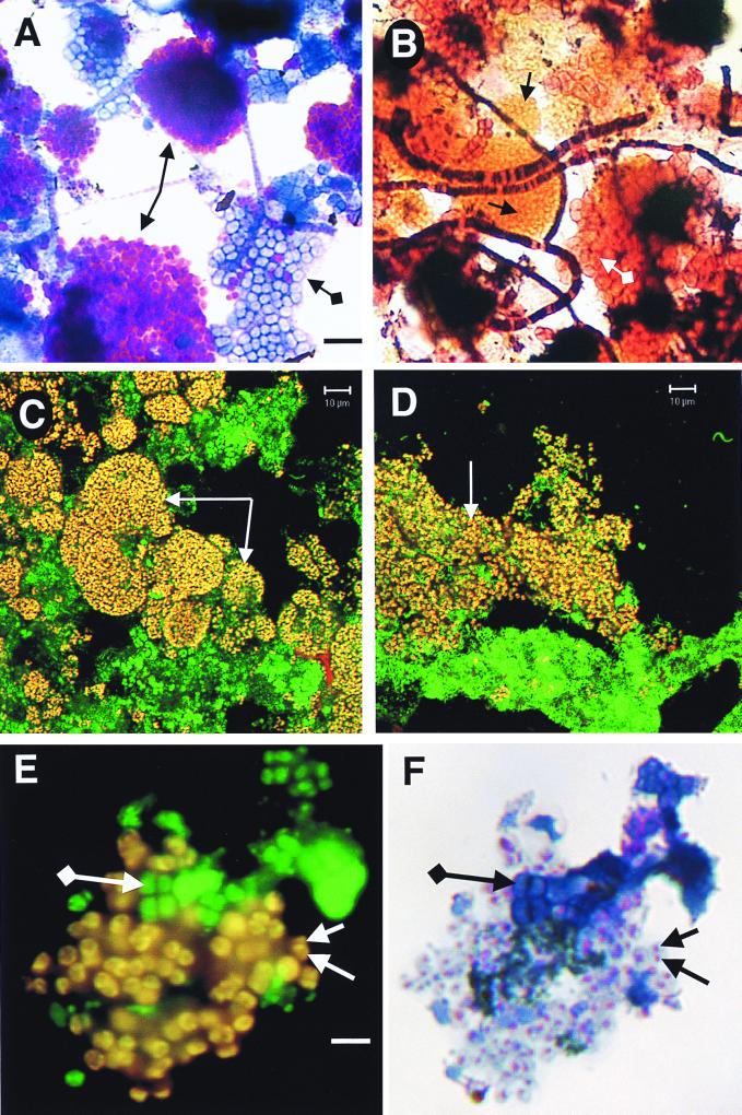
FIG. 2.
Micrographs of mixed liquors from SBRs. (A and B) Bright-field micrographs of GRC sludge as operated according to data in Table 1. (A) Methylene blue stain. Standard-arrowed purple clusters of cells are those containing polyphosphate, while diamond-ended-arrowed blue cells do not contain polyphosphate. The bar is for both panels and is 6 μm. (B) Gram stain. Standard-arrowed orange clusters of cells match the morphology and size of the purple cells in panel A. Diamond-ended-arrowed pink cells match those of the blue cells in panel A. Morphologically identified “Nostocoida limicola” II can be seen as filaments of gram-positive and gram-negative cells. (C and D) Confocal laser scanning micrographs of sludges dual hybridized with EUB338 (25 ng, fluorescein labeled) and a mixture of all three PAO probes (Table 2; each 25 ng, CY3 labeled). Images were collected for fluorescein and CY3 channels, artificially colored, and superimposed. Arrowed yellow cells are the PAOs, since they are dual labeled with EUB338 (green) and PAO (red) probes. The bar for both panels C and D is 10 μm. (C) Mixed liquor from SBR A with operating data as given in Table 1. (D) Lightly sonicated mixed liquor from an EBPR SBR (ca. 10% P in the sludge) operating at 3.5% NaCl from a study of seafood-processing wastewater. Sludge kindly supplied by Nugul Intrasungkha. (E) Epifluorescence micrograph of GRC sludge (Table 1) dual hybridized with EUB338 (25 ng, fluorescein labeled) and PAO651 (Table 2; 25 ng, CY3 labeled). Separate images were collected for fluorescein and CY3 excitation, artificially colored, and superimposed. Standard-arrowed yellow cells are PAOs; diamond-ended-arrowed green-colored cells are other bacteria. The bar is for both panels and is 4 μm. (F) Methylene blue-stained image of the same field as in panel E. Standard-arrowed cells containing purple granules of polyphosphate are the same yellow cells in panel E. Diamond-ended-arrowed blue cells are the same green cells in panel E.

