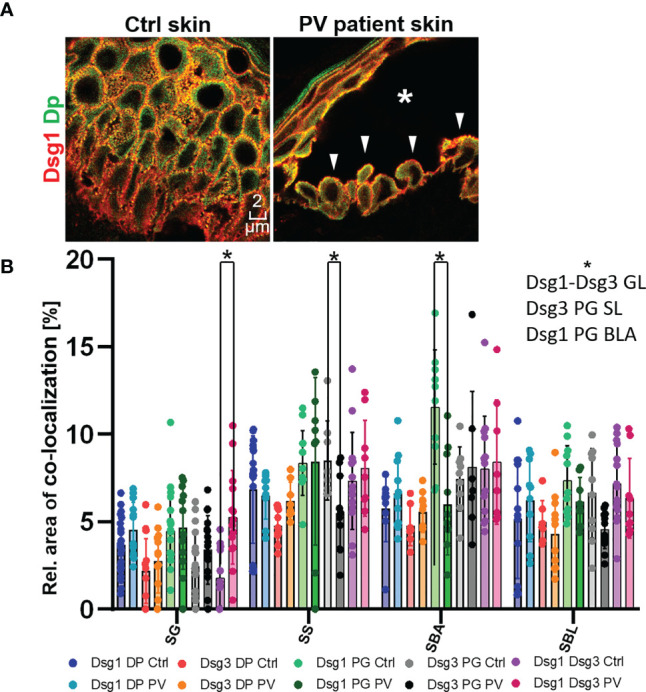
Figure 5.

(A) Confocal microscopy images of a Dsg1 (red) Dp (green) co-staining in control and PV patient skin (featuring a typical PV blister * and tombstoning cells: white pointers). (B) quantification of differences in co-localization of desmosomal proteins in different layers in pemphigus patients compared to control patients N (patients)=3, n (cell borders)=2-7. *Significant difference to the value which is indicated that it is compared to.
