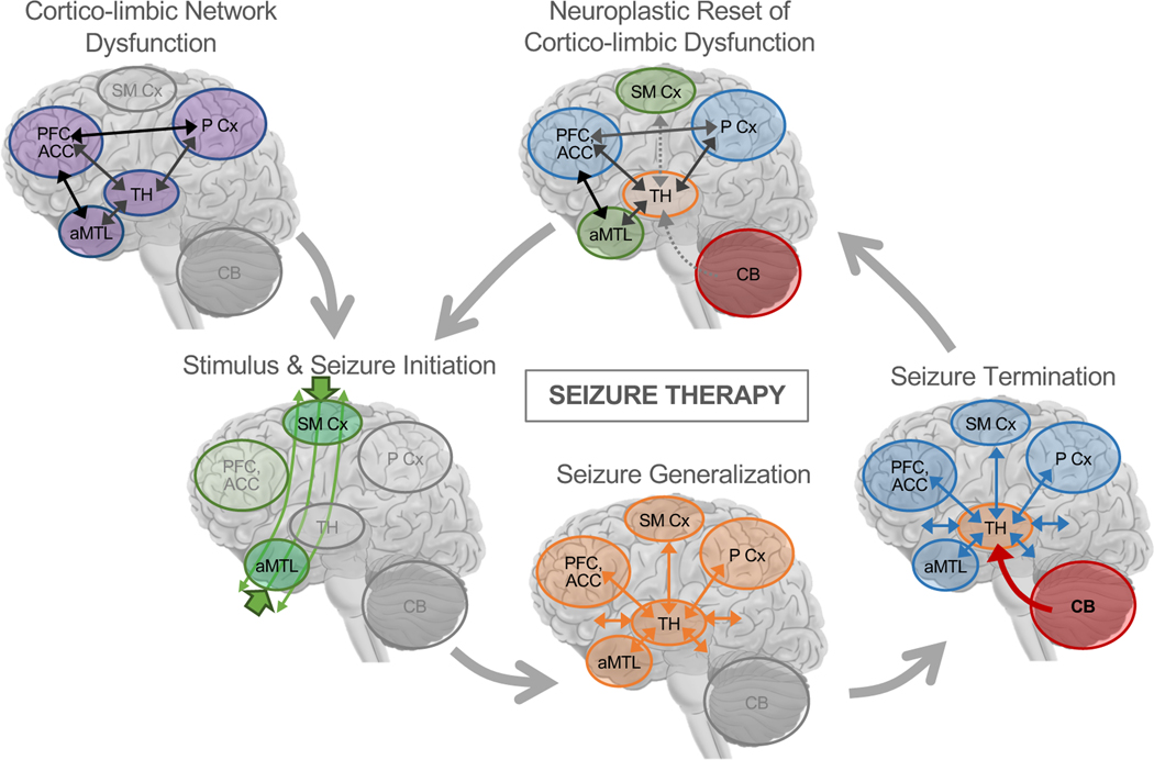
Figure 4.
A Mechanistic Network Model of Therapeutic Seizures. Prior to treatment, patients may have network-level dysfunction in corticolimbic circuits involving anterior medial temporal lobe (aMTL) structures like hippocampus and amygdala, medial and lateral prefrontal cortex (PFC), subgenual and dorsal anterior cingulate cortex (ACC), medial and lateral Parietal Cortex (P Cx), thalamus, and other related structures like ventral striatum previously implicated in neurobiological models and studies of depression (52,53). At the beginning of ECT, the electrical stimulus passes through the head, and localized seizure activity begins near electrode sites. During seizure generalization, synchronized brain activity increases in thalamocortical networks across the brain, perhaps most strongly in regions near electrodes. During seizure termination, cerebellar circuits inhibit generalized seizure activity in thalamo-cortical networks, again perhaps with greater inhibitory control needed in brain regions near electrodes. Over repeated sessions of seizure therapy, these processes constitute a neuroplastic correction or “reset” of corticolimbic dysfunction 1 in depression. When used to target dysfunction in other networks, therapeutic seizure processes in cerebellar-thalamocortical networks may have similar effects on dysfunction in these other networks (e.g., bifrontal ECT to target prefrontal network dysfunction in schizophrenia or depression). Figure adapted with permission [pending] from Leaver et al. 2020 Molecular Psychiatry.

