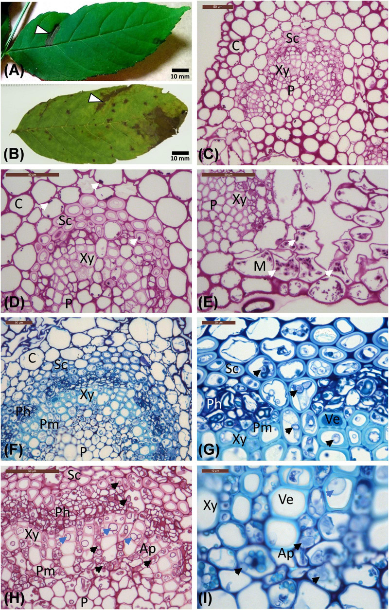
FIGURE 1.
Leaflet images and micrographs of transverse sections of leaflet veins. (A) Leaflet from the Stjørdal site, with a necrotic lesion along one side vein (arrowhead, sample S1) in an otherwise healthy appearing leaflet. (B) Leaflet from the Norderås site with a broad necrotic lesion following one side vein, and several small lesions mostly in leaf blade areas. (C–E) Transverse sections of veins from asymptomatic regions of leaflets from Stjørdal (C,D) and Norderås (E) show the organization of different cell types and the presence of some purple-red stained starch grains (white arrows). (F–I) Transverse sections of veins from lesion areas in leaflets sampled from Stjørdal (F,G) or Norderås (H,I) and showing hyphal presence in sclerenchyma, phloem, axial parenchyma, and perimedullary pith (examples pointed with black arrowheads) and formation of tyloses in vessel elements (examples pointed with blue arrowheads). Note also the appressoria-like swelling upon hyphal spread between two cells of axial parenchyma (I). Ap, Axial parenchyma; C, Cortex; M, Mesophyll; Ph, Phloem; P, Pith; Pm, Perimedullary parenchyma; Sc, Sclerenchyma; Ve, Vessel elements; Xy, xylem.

