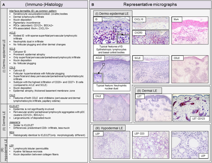
Figure 2.
Overview of typical histopathologic patterns observed in different cutaneous lupus erythematosus (CLE) subtypes. (A) Typical (immuno-)histologic findings of (I) dermo-epidermal lupus erythematosus (LE), (II) dermal LE and (III) hypodermal LE. Dermo-epidermal LE, presenting as interface dermatitis (ID) includes the morphologic variants acute cutaneous LE (ACLE), subacute cutaneous LE (SCLE) and chronic discoid LE (CDLE) among others. Dermal LE consists of intermittent cutaneous LE (ICLE), also named LE tumidus (LET), Jessner-Kanof lymphocyte infiltrate (Jessner's), and reticular erythematous mucinosis (REM), however, some authors consider Jessner's and REM as separate (only lupus-like) entities. Hypodermal LE includes LE profundus (LEP). ID, interface dermatitis; PDCs, plasmacytoid dendritic cells; IFN, interferon. (B) Representative micrographs of different CLE subtypes and selective immunohistochemical features. The typical histopathologic pattern of skin lesions is termed interface dermatitis (ID) and is characterized by epitheliotropic lymphocytes and necroptotic keratinocytes, of which the latter are also called colloid or civatte bodies, at the dermo-epidermal junction. CXCR3+ effector cells are recruited into lesional skin by CXCL10+ expressing keratinocytes. Among these effector cells are CD3+ T lymphocytes, which form the largest immune cell population in LE. The interferon (IFN)-regulated protein MxA reveals a strong expression of IFN in keratinocytes and infiltrating immune cells. ACLE typically features a moderate ID with neutrophilic nuclear dust in the infiltrate. SCLE shows a mild ID with a prominent epidermal atrophy. CDLE features a cell rich ID with a dense perifollicular and perivascular infiltrate and follicular hyperkeratosis and plugging. ICLE/LET presents with a patchy dermal infiltrate and large amounts of deposited mucin. In LEP, a lymphocytic lobular panniculitis can be observed.

