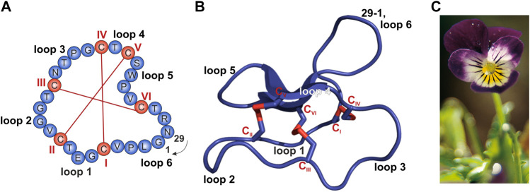
FIGURE 1.
Sequence and structure of the prototypic cyclotide kalata B1 and photograph of Viola tricolor. (A,B) The cyclized peptide kalata B1 (PDB 1NB1) comprises 29 residues (blue) with six cysteine residues (C; red; Roman numerals) which form three disulfide bonds establishing the typical cyclic cystine knot (CKK) motif. The cyclization point between residue 1 and 29 is indicated. (A) Amino acid sequence obtained from the CyBase databank (Wang et al., 2007). (B) Cartoon of prototypical cyclotide structure of kalata B1 prepared with PyMol. (C) Photograph of Viola tricolor, kindly provided by C. Gründemann, courtesy of Weleda AG, Schwäbisch-Gmünd, Germany.

