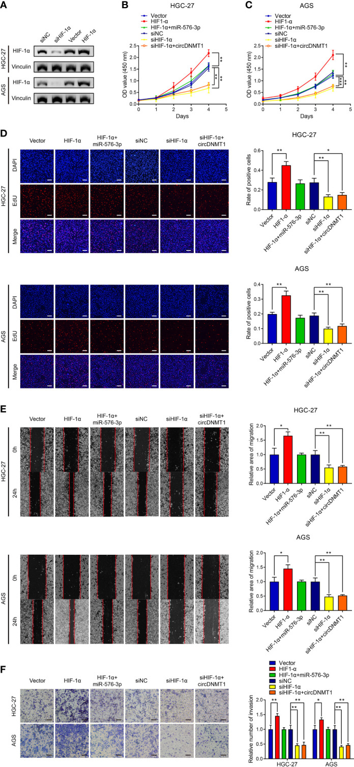
Figure 6.

circDNMT1 promotes GC malignancy by targeting miR-576-3p/HIF-1α axis. (A) The WB analysis to show HIF-1α expression in HGC-27 and AGS cells transfected with NC shRNA or HIF-1α siRNA, or carrying lentivirus with vector or HIF-1α overexpression plasmids. (B, C) The CCK-8 assay to show proliferation of HGC-27 (B) and AGS (C) cells. They could be divided into two groups: i) The cells stably carrying lentivirus with vector or HIF-1α overexpression plasmids and transfected with NC or miR-576-3p mimics; ii) The cells transfected with NC shRNA or HIF-1α siRNA and carrying lentivirus with vector or circDNMT1 overexpression plasmids. (D) The EdU assay to show proliferation of cells as in (B, C). Scale bar: 100 μm. The histogram is displayed on the right. (E) The wound healing assay to show migration of cells as in (B, C). The histogram is displayed on the right. (F) The transwell assay to show invasion of cells as in (B, C). The histogram is displayed on the right. Scale bar: 100 μm. Data were presented as means ± SD. *P < 0.05, **P < 0.01, ***P < 0.001.
