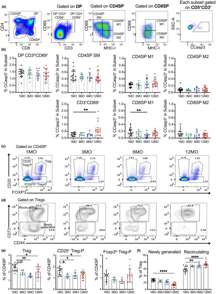
FIGURE 5.

Aging associated changes in central tolerance of polyclonal thymocytes. (a) Representative flow cytometry plots showing identification of thymocyte subsets undergoing negative selection. Post‐positive selection DP thymocytes were subdivided into DP CD3loCD69+ and DP CD3+CD69+ stages; CD4SP and CD8SP cells were divided into semi‐mature (SM), mature1 (M1) and mature2 (M2) stages, as indicated. Cells were gated on CD5+ CD3+ cells to restrict analysis to thymocytes that had undergone TCR signaling, and cleaved caspase 3 (CCasp3) expression identified cells undergoing clonal deletion in each subset. (b) Quantification of the frequency of CCasp3+ cells in thymocyte subsets from mice at 1, 3, 6 and 12 MO of age. (c‐d) Representative flow cytometry plots of (c) CD25 and FOXP3 to distinguish Treg‐P and Tregs and (d) CD73 on Tregs to distinguish newly generated (CD73−) from recirculating (CD73+) cells in thymuses from mice at 1, 3, 6 and 12 MO of age. (e) Percentage of Tregs, CD25+ Treg‐Ps, and FOXP3lo Treg‐Ps in the CD4SP compartment at the indicated ages. (f) Percentage of newly generated versus recirculating Tregs at the indicated ages. (b, e‐f) Plots show mean ± SEM of nine to fifteen thymuses per age. Symbols represent individual thymuses. Analyzed by one‐way ANOVA with Tukey's test for multiple comparisons, p‐values: * <0.05, ** <0.01, *** <0.001, **** <0.0001
