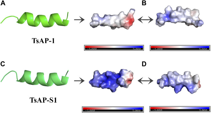
FIGURE 3.
Antimicrobial peptides visualized using PyMol. (A,C) Peptide tridimensional view of TsAP-1 and TsAP-S1, respectively. (B,D) Different angles of the electrostatic surface created through Adaptive Poisson–Boltzmann solver (APBS). The negatively charged surface is shown in red, the positively charged surface is shown in blue, and the neutrally charged surface is shown in white.

