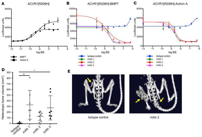
Figure 1. Anti-ACVR1 antibodies block BMP7 and activin A signaling in HEK293.ACVR1[R206H] cells but increase heterotopic bone formation in FOP mice.
Activin A and BMP7 dose response was evaluated in stable pools of HEK293/BRE-luciferase reporter cells overexpressing ACVR1[R206H] (A). HEK293/BRE-luciferase reporter cells overexpressing ACVR1[R206H] were treated with a fixed concentration (2 nM) of BMP7 (B) or activin A (C). Anti-ACVR1 antibodies inhibited Smad1/5/8 phosphorylation induced by BMP7 or activin A (B and C). Data show the mean (n = 4) ± SEM. Three biological replicates were performed for the in vitro signaling assays. (D) Acvr1[R206H]FlEx/+; GT(ROSA26)SorCreERT2/+ mice were injected with tamoxifen to initiate the model and concurrently injected with anti-ACVR1 antibodies or isotype control antibody at 10 mg/kg weekly (n = 7–8/group). Total heterotopic bone lesion volume was measured 4 weeks after initiation. Data show the mean ± SD. *P < 0.05, **P < 0.01 by 1-way ANOVA with Dunnett’s multiple-comparison test. (E) Representative μCT images of FOP mice [Acvr1[R206H]FlEx/+; GT(ROSA26)SorCreERT2/+, after tamoxifen] treated with anti-ACVR1 antibody or isotype control antibody. Yellow arrows indicate the positions of heterotopic bone lesions.

