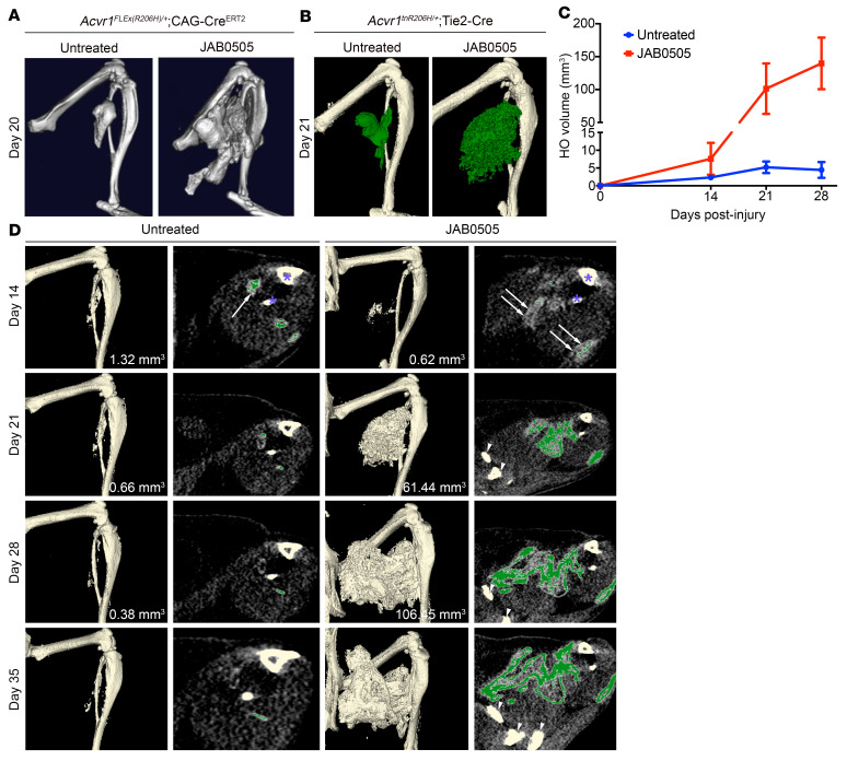
Figure 2. JAB0505 profoundly exacerbates HO and extends the period of lesional growth in FOP mice.
(A) Representative μCT images of HO in Acvr1FLEx(R206H)/+; CAG-CreERT2 mice 20 days after cardiotoxin-induced injury of the gastrocnemius muscle (Untreated, n = 3; JAB0505, n = 4). (B) Representative μCT images of HO (pseudocolored green) in Acvr1tnR206H/+; Tie2-Cre mice 21 days after pinch injury of the gastrocnemius muscle (Untreated, n = 11; JAB0505, n = 10). (C) Quantification of HO volumes as a function of time after muscle pinch injury of Acvr1tnR206H/+; Tie2-Cre mice. Untreated, n = 11; JAB0505 (10 mg/kg), n = 6. Error bars represent ±SEM. ****P ≤ 0.0001 by 2-way ANOVA with Sidak’s multiple-comparison test. (D) Paired single transverse slice and 3D reconstructed μCT images of the distal hind limb of Acvr1tnR206H/+; Tie2-Cre mice at the indicated times after hind limb muscle pinch injury with and without administration of JAB0505. Mineralized bone in the slice images is pseudocolored green. Radio-opaque lesional tissue below the threshold set for quantification of mineralized bone (white arrows in day 14 slices) is extensive at day 14 in JAB0505-treated mice. Mineralized bone in day 14 slices is barely visible at this magnification. HO volumes are given for images prior to day 35. The tibia and fibula are labeled with asterisks in the day 14 slices. Pelvic bones present in day 21, 28, and 35 slices of JAB0505-treated mice are denoted with arrowheads. To avoid confusion with HO, the baculum present in some images was removed by segmentation.

