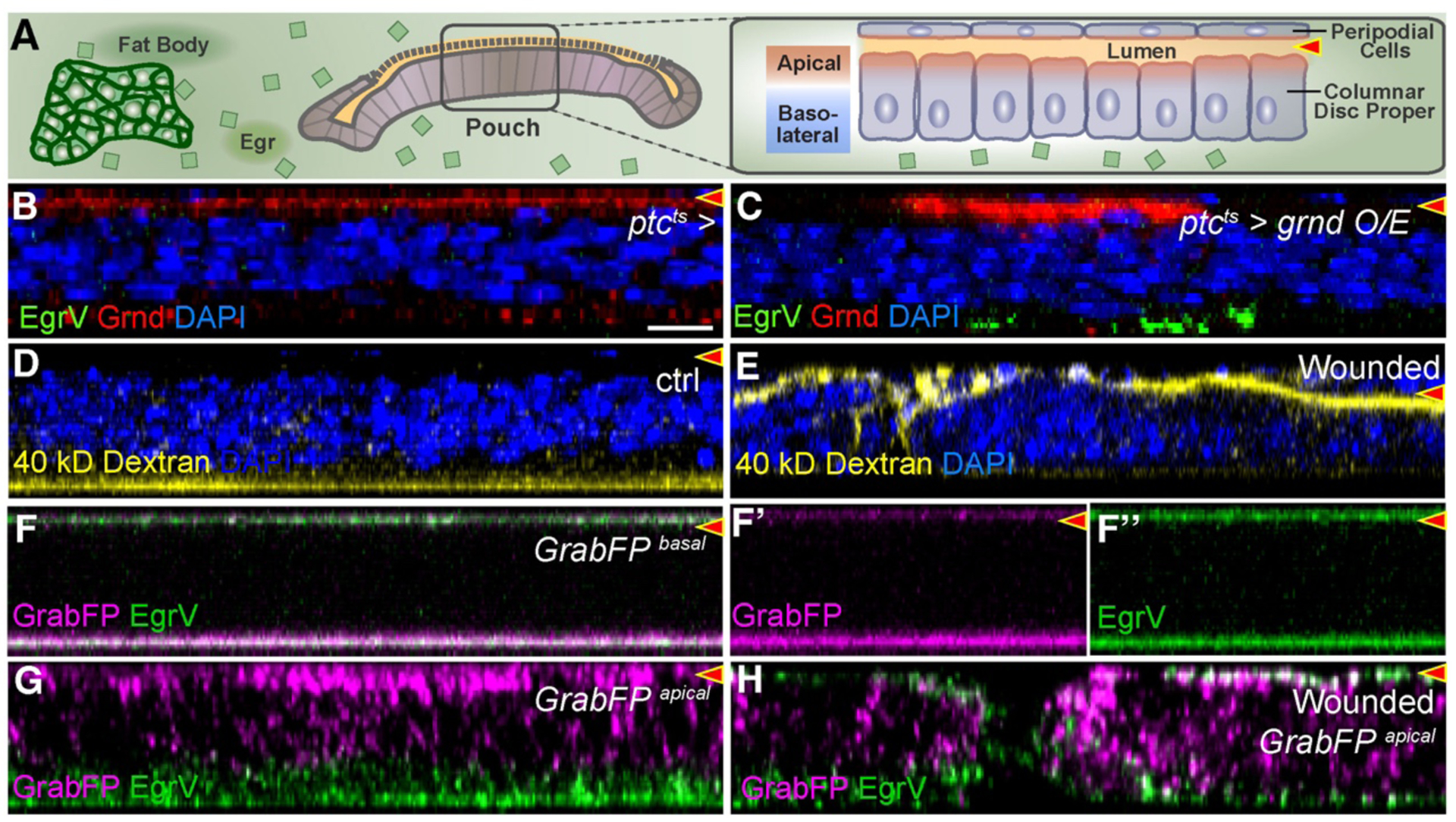
Fig. 3. Egr binds basolaterally to polarity-deficient cells.

X-Z cross-sections show disc proper below and peripodium above. Lumen is indicated by red arrowhead.
(A): Diagram showing relationship of disc epithelial barrier to hemolymph. Egr secreted by fat body bathes the basolateral surface but is excluded from apical surface and lumen.
(B-C): Grnd is apically localized (B), even when overexpressed (C), but bound EgrV is exclusively basolateral.
(D-E): Dextran in media is excluded from lumen of intact discs (D) but can enter wounded discs (E).
(F): Basolateral GrabFP binds strongly at basal surface to EgrV produced by fat bodies. Signal at top is peripodial basal surface. F’ and F” show left half of F.
(G-H): Apical GrabFP binds EgrV only at the basolateral surface (G), but wounding enables strong apical binding of EgrV as well (H).
Scale bar: 10 μm in B
