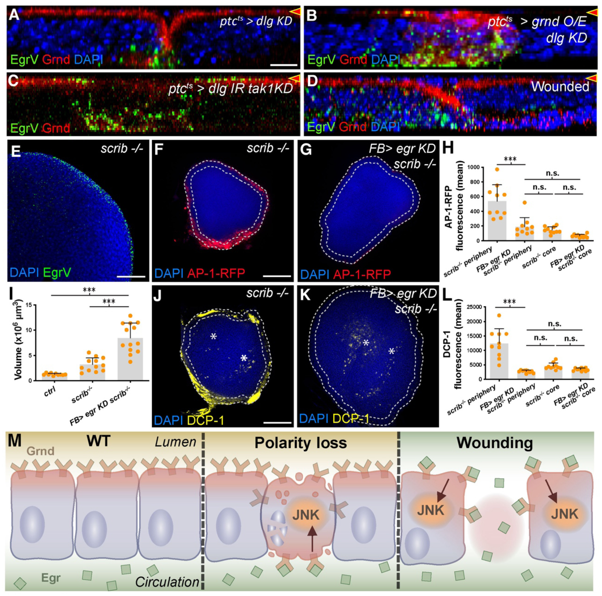
Fig. 4. Mispolarization of Grnd permits Egr binding and cell elimination.

(A-D): X-Z sections as in Fig. 3. dlg-depleted cells mislocalize Grnd basolaterally (A), evident especially when Grnd is overexpressed (B). EgrV binds basally to dlg-depleted cells. Co-depletion of dlg and tak1 (C) blocks cell elimination but polarity defects remain. Mispolarized Grnd binds EgrV at basal surface. Wounded discs (D) show altered Grnd localization and basal EgrV binding.
(E-L): X-Y sections. Wing discs containing only scrib mutant cells bind Egr preferentially at periphery (E). JNK signaling (F) is elevated in periphery compared to core and is dependent on circulating Egr (G, quantitated in H). scrib discs grow larger when circulating Egr is depleted (I), and peripheral apoptosis (J, DCP-1) is reduced (K, asterisks indicate DCP-1+ cells in core; quantitated in L).
(M): Model for role of polarized segregation of TNF ligand and receptor in epithelial homeostasis.
Scale bar: 10 μm in A, 50 μm in E, 100 μm in F, and J.
