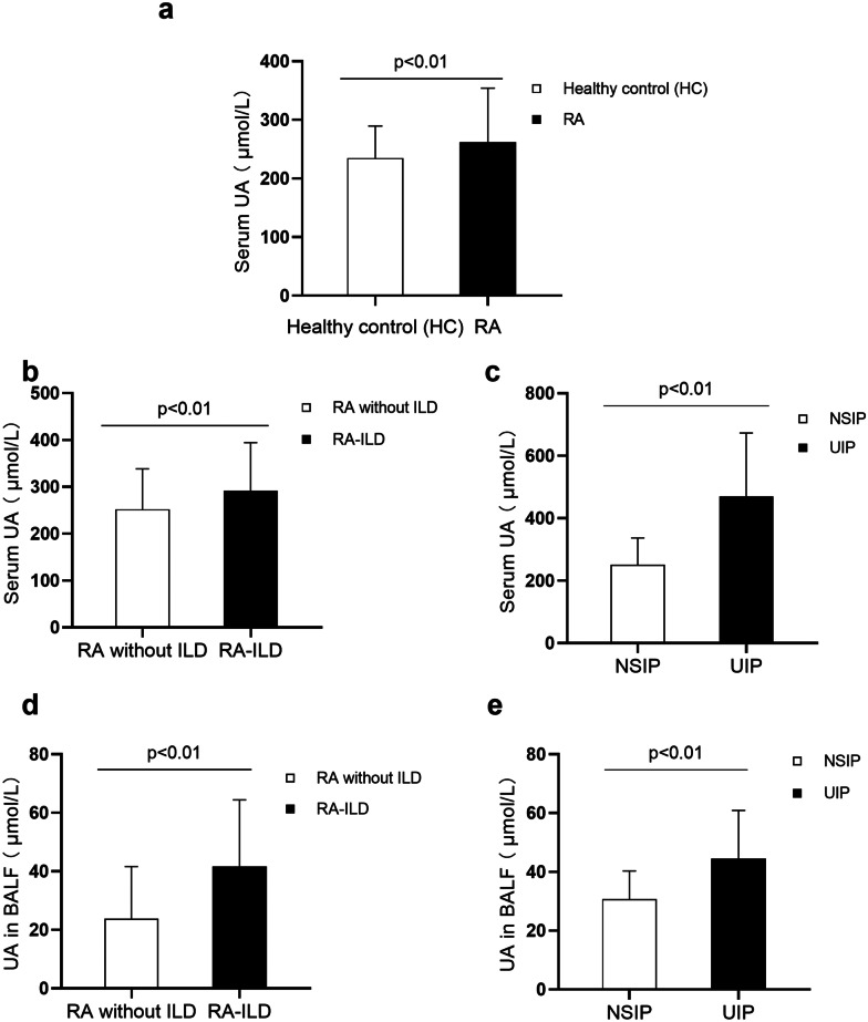
Fig. 1.
The level of UA was measured in patients with RA-ILD. The level of UA in the serum of patients with RA (n = 266) and healthy control (HC) (n = 138). Statistical difference was detected between patients with RA and HC (p < 0.01) a. Comparison of serum UA levels between non-ILD and RA-ILD, as well as NSIP and UIP. The higher serum UA levels were observed in patients with RA-ILD (n = 162) relative to RA patients without ILD (n = 104) (p < 0.01) b. Statistical difference of serum UA was also detected between UIP (n = 76) and NSIP (n = 86) patterns of RA-ILD (p < 0.01) c. The UA level in BALF of patients with RA (n = 97) was also measured. Compared with RA patients without ILD (n = 39), the level of UA in patients with RA-ILD (n = 58) was significantly higher (p < 0.01) d. And, a marked increase of the level of UA was observed in BALF in patients with RA-UIP (n = 37) relative to RA patients with NSIP pattern (n = 21) (p < 0.01) e.

