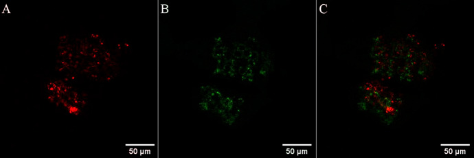
Fig. 1.
Confocal imaging of a single live human pancreatic cancer (HPAC) spheroid treated with an Os(II) polyarginine probe, [Os-(R4)2]10+ at 100 μM/48 h. Using a 490 nm white light laser for excitation, emission was collected between A 650 and 800 nm; Os(II) channel and B 500–570 nm; auto-fluorescence window. C Os(II)/autofluorescence channel overlay. Reprinted (adapted) with permission from Ref. [10] (https://pubs.acs.org/doi/10.1021/acs.inorgchem.1c00769). Further permissions related to the material excerpted should be directed to the ACS

