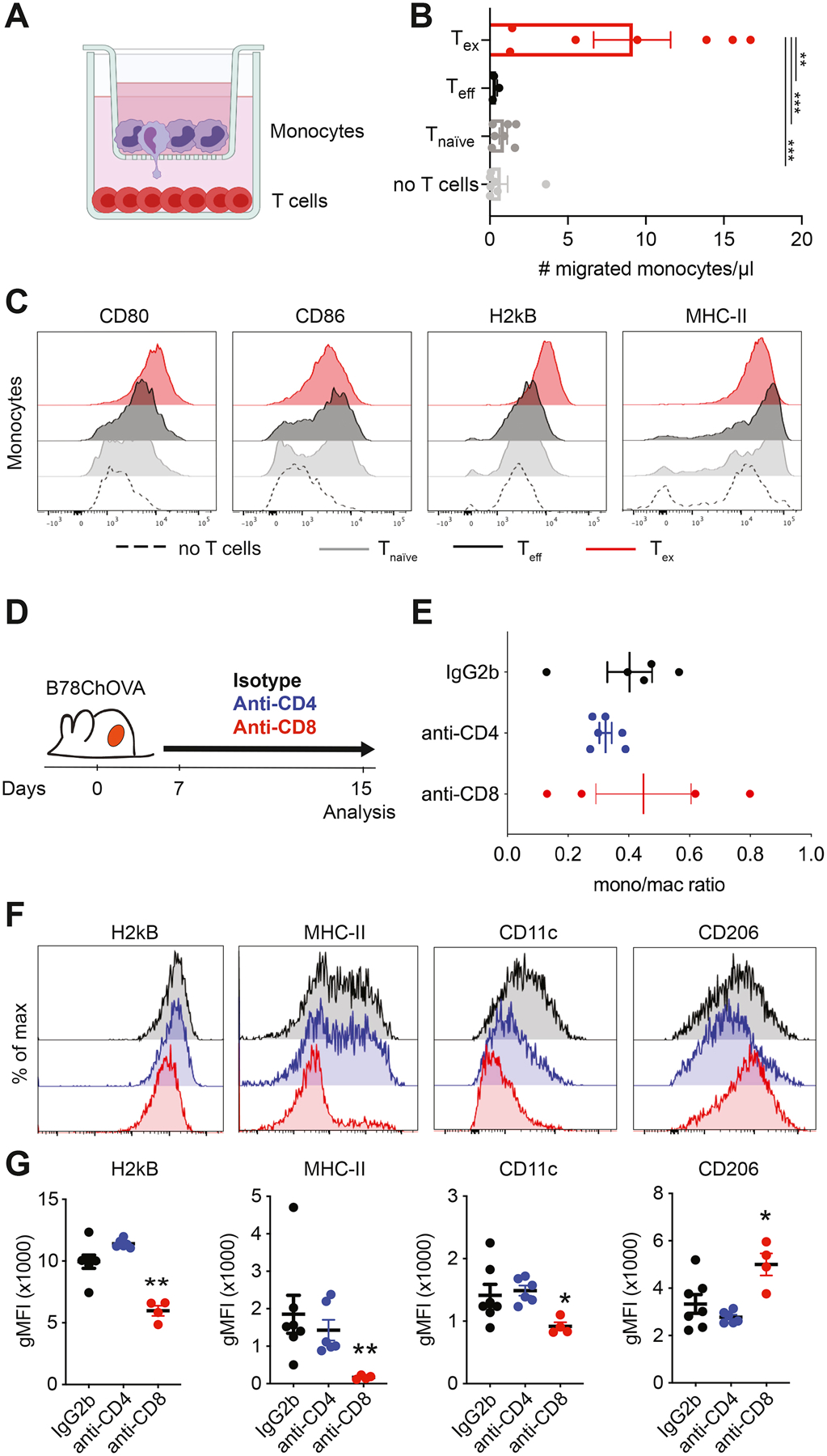
Figure 3. Exhausted CD8+ T cells recruit monocytes to the TME and shape macrophage phenotype.

A) Experimental setup of in vitro recruitment assay. Bone marrow-derived monocytes are cultured on transwell inserts (5μm pore size) and T cells (OT-I Tnaïve, Teff and Tex) are plated in the bottom well. B) Quantification of recruited monocytes after 24 hours. Data combined from two independent experiments. Statistical significance was determined by one-way ANOVA with Holm-Sidak’s multiple testing correction. C) Representative histograms of expression of surface markers on monocytes after 48 hours of co-culture with Tnaïve, Teff and Tex cells. D) Experimental set-up of in vivo CD4+ and CD8+ T cell depletion in B78ChOVA-bearing mice. Treatment with anti-CD4/CD8 antibodies or isotype was initiated 7 days after tumor inoculation and continued until mice were sacrificed. E-F) Monocyte/macrophage ratio of the proportion of Ly6Chi monocytes and F4/80+ macrophages (gated of CD45+ cells) in the B78ChOVA TME after isotype, anti-CD4 and anti-CD8 treatment. F-G) Representative histograms (F) and quantification (G) of H2kb, MHC-II, CD11c and CD206 expression on CD11b+F4/80+ macrophages in B78ChOVA tumors after isotype, anti-CD4 and anti-CD8 treatment. Statistical significance was determined using the Mann-Whitney U test. All data are mean ± SEM. * p < 0.05, ** p < 0.01, *** p < 0.001. See also Figure S3.
