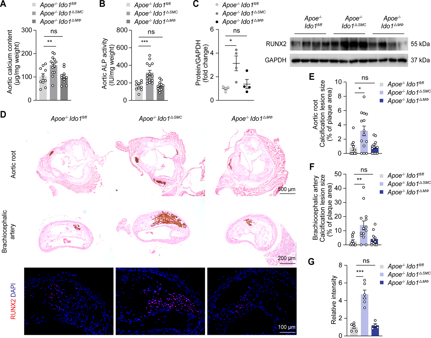
Figure 2. VSMCs-specific IDO1 deficiency promotes calcification and RUNX2 expression.

Apoe−/− Ido1fl/fl (n = 10), Apoe−/− Ido1fl/fl Myh11Cre (Apoe−/− Ido1△SMC, n = 14), and Apoe−/− Ido1fl/fl lysMCre (Apoe−/− Ido1△MΦ, n = 11–12) mice were fed with Western diet for 24 weeks.
A and B, Biochemical measurement of aortic calcium content (A) and ALP activity (B) for indicated mice.
C, Western blot analysis of RUNX2 expression in aorta lysates from indicated mice (n = 4).
D, Representative von Kossa staining and immunofluorescence staining of RUNX2 in aortic root and brachiocephalic artery tissue sections from indicated mice. Scale bars: 500 μm for von Kossa staining of aortic root; 200 μm for von Kossa staining of brachiocephalic artery; 100 μm for immunofluorescence staining.
E-G, Quantification of percentage of calcification lesions within plaque areas in aortic root (E) and brachiocephalic artery tissue sections (F) from Apoe−/− Ido1fl/fl, Apoe−/− Ido1△SMC, and Apoe−/− Ido1△MΦ mice, and RUNX2 immunofluorescence staining (G) in brachiocephalic artery tissue sections from indicated mice (n = 4–6 for G).
Results are presented as mean ± SEM. P values are assessed using one-way ANOVA with Dunnett’s post hoc test for A-C and G, Welch’s ANOVA with Dunnett’s post hoc test for E and F. *P < 0.05, **P < 0.01, ***P < 0.001, ns indicates P > 0.05.
