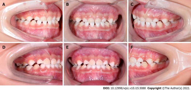
Figure 3.
Intraoral photographs under treatment. A: Lateral view of right side of intraoral pictures of the 4th aligner; B: Frontal view of intraoral pictures of the 4th aligner; C: Lateral view of left side of intraoral pictures of the 4th aligner; D: Lateral view of right side of intraoral pictures of the 13th aligner; E: Intraoral pictures of the 13th aligner; F: Lateral view of left side of intraoral pictures of the 13th aligner.

